synchrotron radiation x-ray phase contrasting computed tomography and experimental method thereof

A phase-contrast imaging and phase-contrast technology, applied in computerized tomography scanners, echo tomography, etc., can solve the problem of not being able to obtain three-dimensional structures

Inactive Publication Date: 2007-05-23
INST OF HIGH ENERGY PHYSICS CHINESE ACADEMY OF SCI
View PDF0 Cites 17 Cited by
  • Summary
  • Abstract
  • Description
  • Claims
  • Application Information

AI Technical Summary

Problems solved by technology

[0004] Although both theory and experiments have shown that X-ray phase contrast imaging can achieve high contrast and resolution, it is still a two-dimensional imaging, which is the superimposition of the projected images of the sample at various levels perpendicular to the beam direction, and the sample cannot be obtained. three-dimensional structure

Method used


Image

  • synchrotron radiation x-ray phase contrasting computed tomography and experimental method thereof
  • synchrotron radiation x-ray phase contrasting computed tomography and experimental method thereof
  • synchrotron radiation x-ray phase contrasting computed tomography and experimental method thereof

Examples


Embodiment Construction

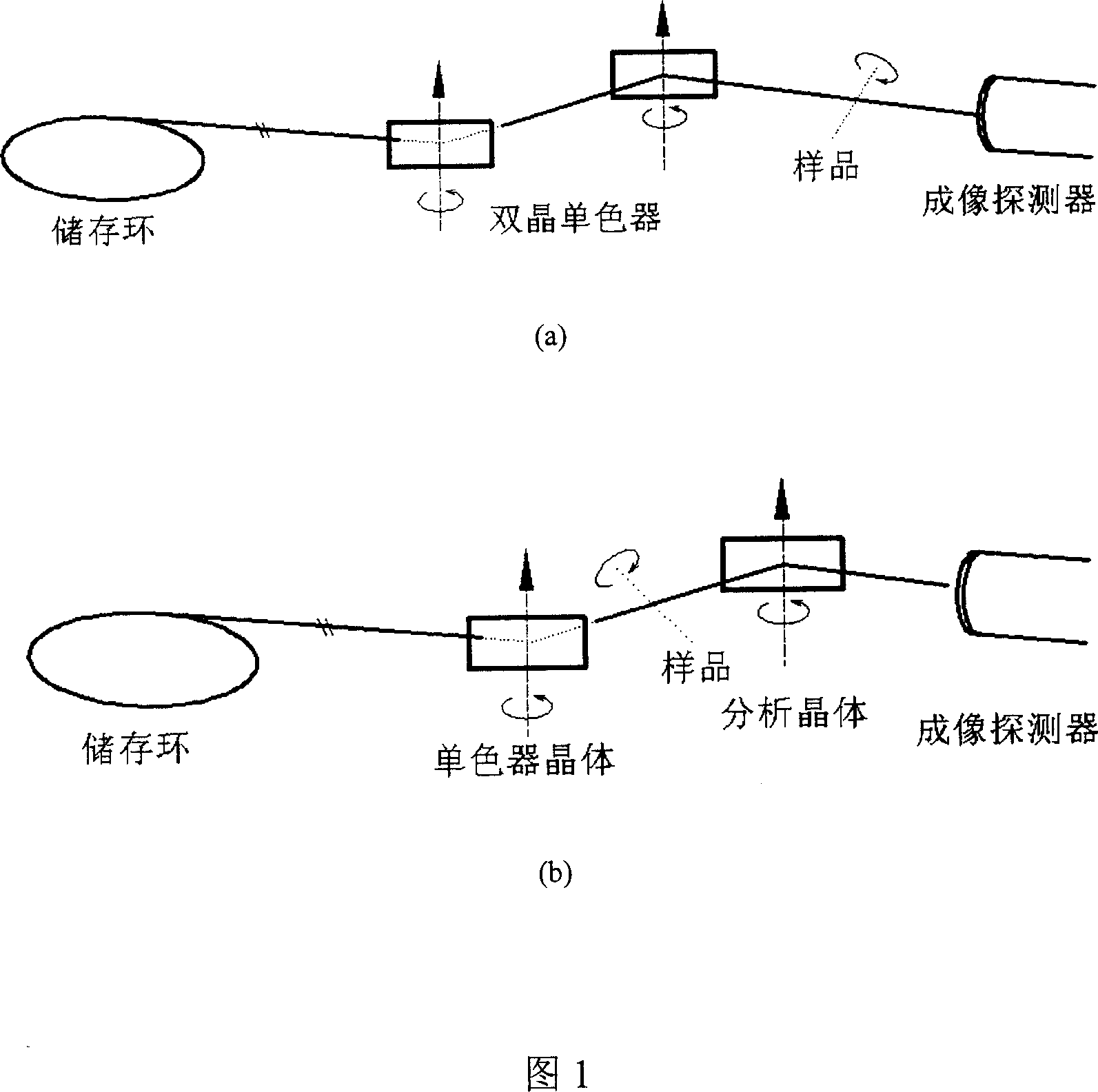
[0036] Figure 1 is a schematic diagram of the optical paths of two imaging modes widely used in phase contrast imaging. Among them, Fig. 1(a) is the optical path diagram of the coaxial phase contrast CT imaging experiment, and Fig. 1(b) is the optical path diagram of the diffraction-enhanced phase contrast CT imaging experiment.

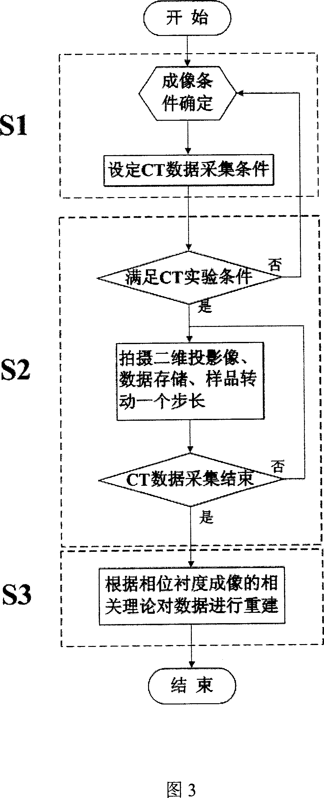
[0037] In synchrotron radiation phase-contrast imaging experiments, since the light source is fixed, subsequent experimental devices must be adjusted according to the position and direction of the light source. Therefore, in phase-contrast CT imaging CT experiments, the acquisition of projection data can only be achieved through the rotation of the experimental sample. Not the rotation of the light source to do it.

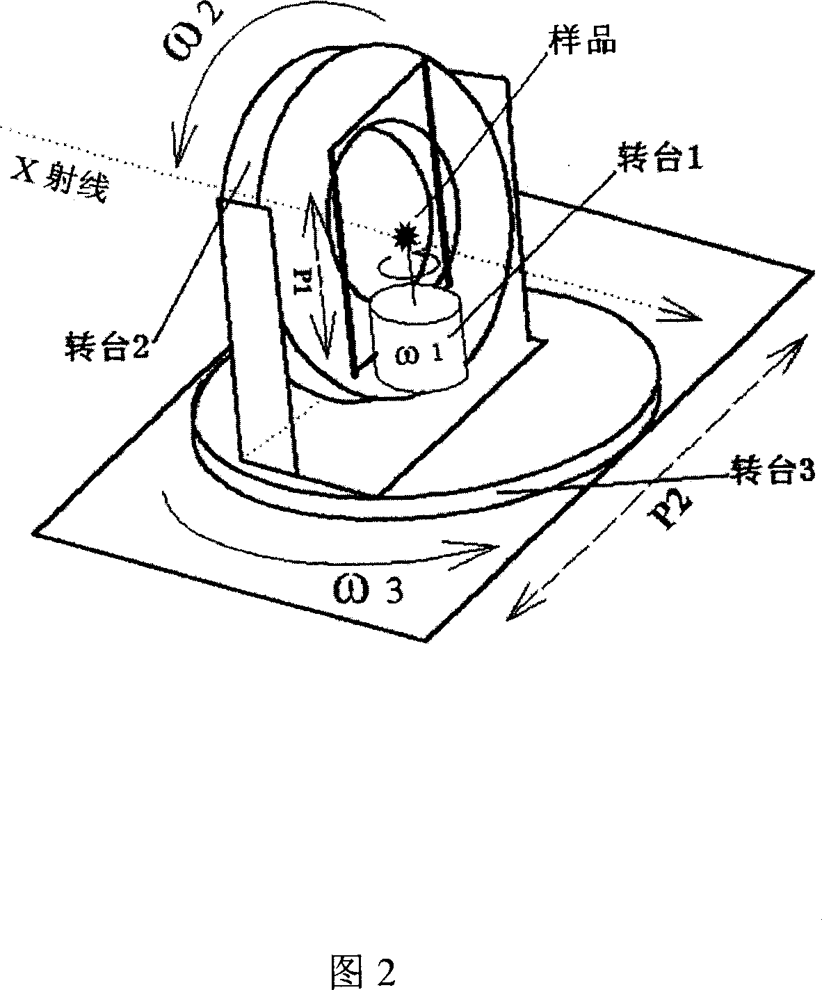
[0038] In the synchrotron radiation X-ray phase contrast CT imaging device of the present invention, the device includes a monochromator crystal, a sample turntable, an analysis crystal, an ionization chamber, and an imaging detector. In...


PUM


Abstract

The invention relates to a computer tomography imaging technique, especially a synchronous radiation X-ray phase contrast CT imager and test method, wherein it is formed by monochromator crystal, sample table, analysis crystal, ionize room, and image detector; the sample table is formed by three rotations and two translations; the method comprises that S1 , finding the image conditions of phase contrasts in different image modes; S2, obtaining phase contrast CT test data; S3, rebuilding the test data.

Description

technical field [0001] The invention relates to the technical field of computer tomography, in particular to a synchrotron radiation X-ray phase contrast CT imaging device and an experimental method. Background technique [0002] Since Roentgen discovered X-rays in 1895, X-ray fluoroscopy imaging based on absorption contrast has become an important detection method in the fields of medical diagnosis and material research because it can obtain the internal structure information of samples non-destructively. . Especially after the invention of X-ray CT (computed tomography) by A.M.Cormack and G.N.Hounsfield and others in the middle and late 20th century, CT has become an essential means of medical diagnosis. But whether it is X-ray fluoroscopic imaging or traditional CT, the imaging contrast is based on the difference in the absorption of X-rays by the sample. For biological soft tissues with light elements as the main components, since the absorption of light element materi...

Claims


Application Information

Patent Timeline
Patent Type & Authority Applications(China)
IPC IPC(8): A61B6/03
Inventor 吴自玉袁清习王寯越朱佩平黄万霞舒航
Owner INST OF HIGH ENERGY PHYSICS CHINESE ACADEMY OF SCI
Why Eureka
Try Eureka