

System for harmonizing medical image presentation
a medical image and presentation technology, applied in image enhancement, instruments, applications, etc., can solve the problem that the normal diagnostic accuracy of hicv data is inferior
- Summary
- Abstract
- Description
- Claims
- Application Information
AI Technical Summary
Benefits of technology
Problems solved by technology
Method used
Image

Examples
Embodiment Construction
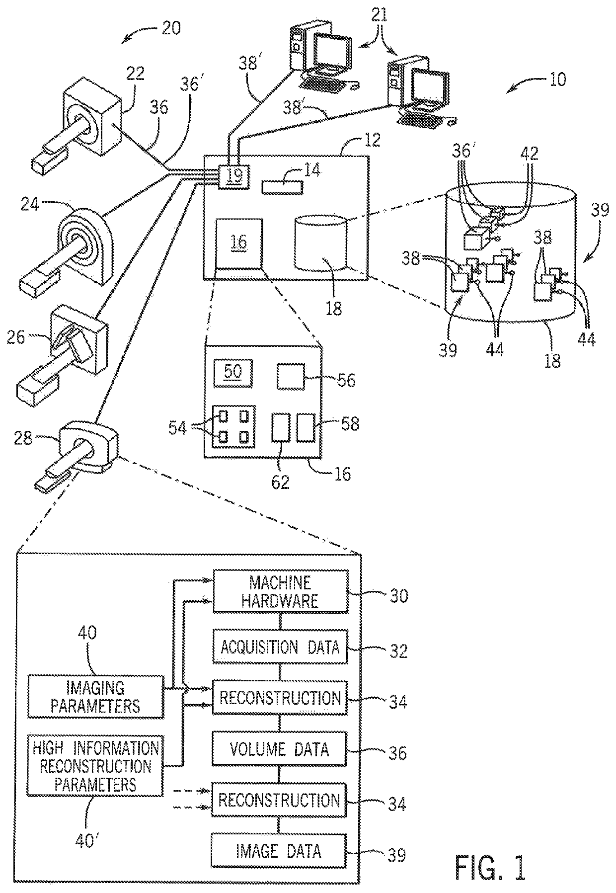
[0059]Referring now to FIG. 1, a medical image harmonizing system 10 per the present invention may provide for a presentation server 12 including an electronic processor 14 communicating with a memory 16 and a database 18. The presentation server 12 may provide for a standard general-purpose computer architecture used for data servers and the like.
[0060]In this regard, the presentation server 12 may provide for a network interface 19 communicating with multiple medical imaging machines 20, for example, the medical imaging machines 20 including but not limited to an MRI machine 22, a CT machine 24, a SPECT machine 26, and a PET machine 28, each of types well known in the art.
[0061]Each of these medical imaging machines 20 may provide for machine-specific acquisition software and hardware 30 for acquiring data from a volume of patient tissue of the patient being scanned by the particular medical imaging machine 20. That hardware 30 may include, for example, a rotating gantry with x-ra...
PUM
 Abstract
Description
Claims
Application Information