System for Harmonizing Medical Image Presentation

Active Publication Date: 2020-07-30
WISCONSIN ALUMNI RES FOUND
View PDF2 Cites 1 Cited by
  • Summary
  • Abstract
  • Description
  • Claims
  • Application Information

AI Technical Summary

Benefits of technology

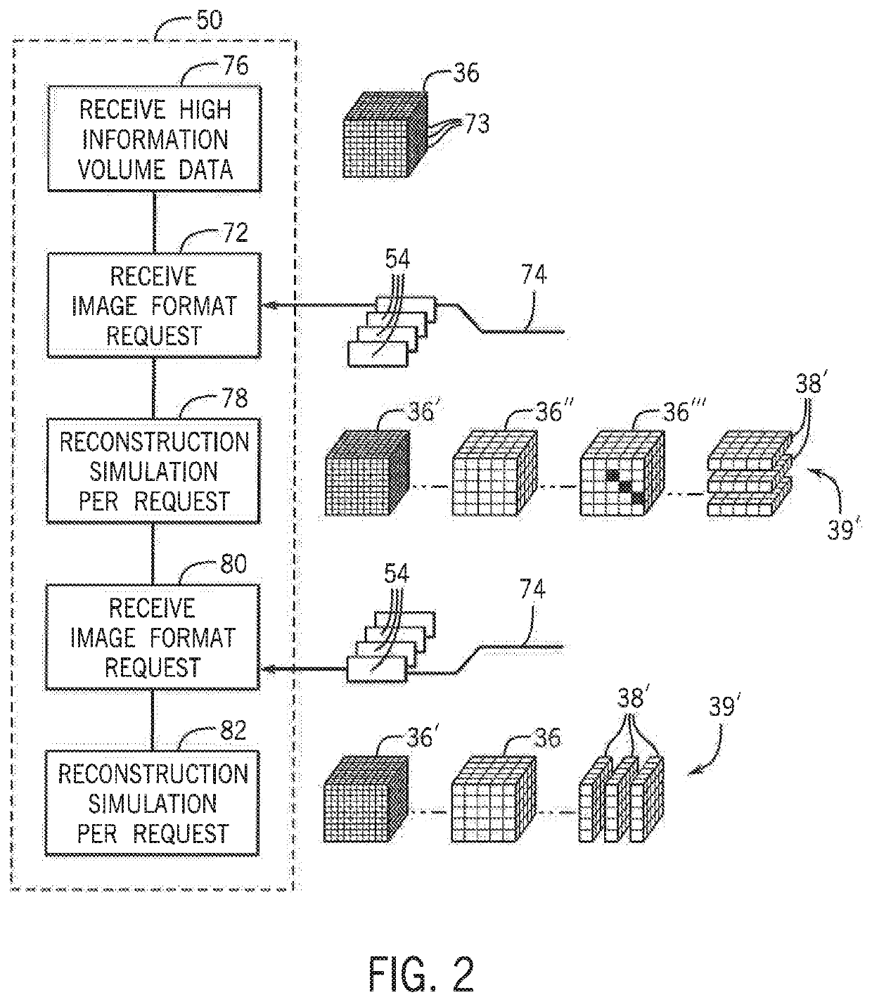
[0014]The present inventor has recognized that significant medical image features normally adjusted during reconstruction can in fact be adjusted after reconstruction eliminating the need to have access to the reconstruction engine of the medical imaging device. This ability to adjust the images after reconstruction is possible by performing an initial reconstruction to produce “high information co

Problems solved by technology

HICV data is normally diagnostically inferior to image data produced by a reconstruction engine for a human diagnostician; nevertheless, HICV data can be

Method used


Image

  • System for Harmonizing Medical Image Presentation
  • System for Harmonizing Medical Image Presentation
  • System for Harmonizing Medical Image Presentation

Examples


Example

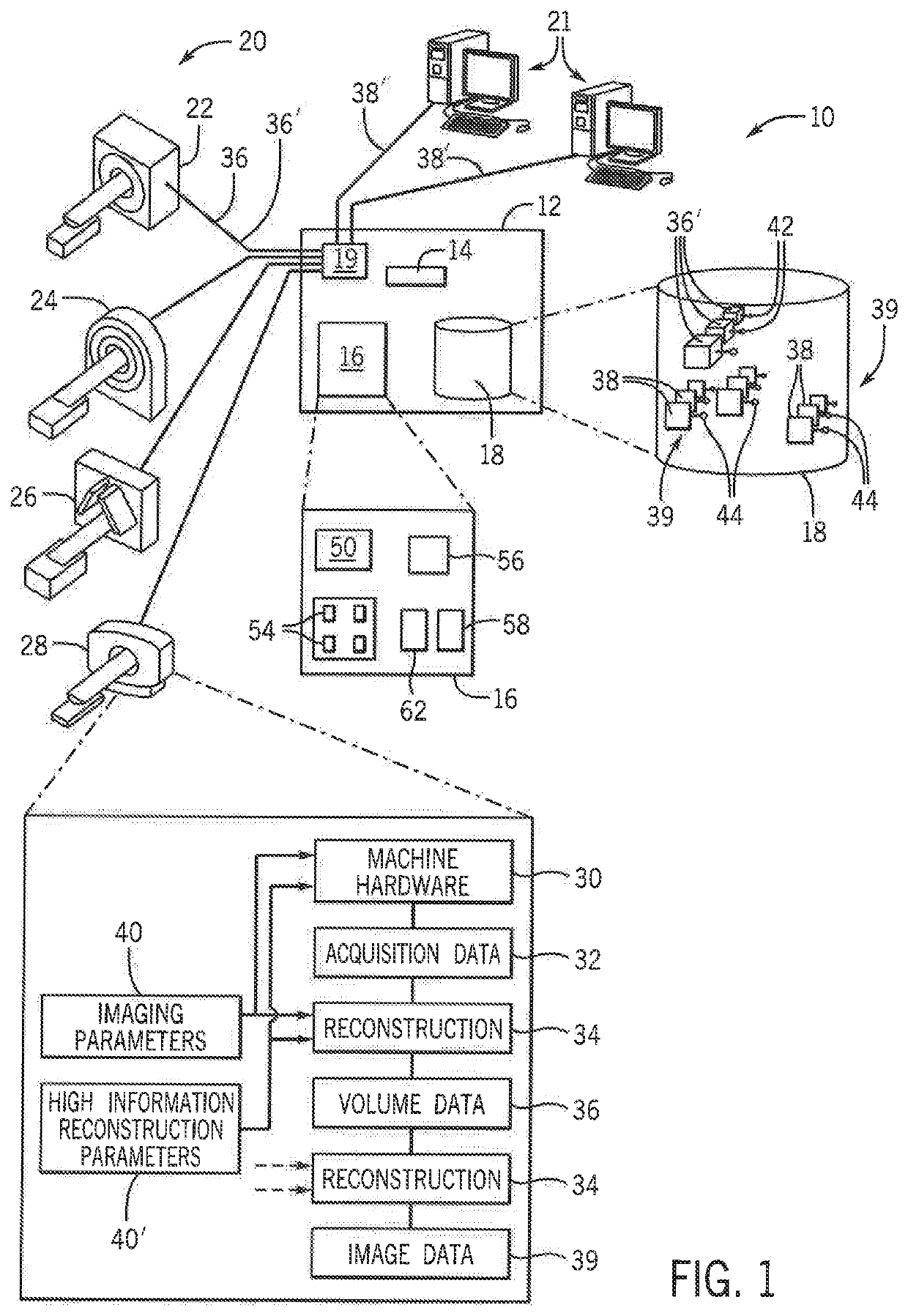
[0059]Referring now to FIG. 1, a medical image harmonizing system 10 per the present invention may provide for a presentation server 12 including an electronic processor 14 communicating with a memory 16 and a database 18. The presentation server 12 may provide for a standard general-purpose computer architecture used for data servers and the like.

[0060]In this regard, the presentation server 12 may provide for a network interface 19 communicating with multiple medical imaging machines 20, for example, the medical imaging machines 20 including but not limited to an MRI machine 22, a CT machine 24, a SPECT machine 26, and a PET machine 28, each of types well known in the art.

[0061]Each of these medical imaging machines 20 may provide for machine-specific acquisition software and hardware 30 for acquiring data from a volume of patient tissue of the patient being scanned by the particular medical imaging machine 20. That hardware 30 may include, for example, a rotating gantry with x-ra...


PUM


Abstract

A system for processing medical image data stores high information content volumetric (HICV) data reconstructed from different medical imaging devices. Images normally requiring differing image imaging parameters can instead be created by processing of the HICV data, importantly without access to the original reconstruction algorithms or machine. The invention also allows different medical images to be harmonized for ready comparison or according to a desired image sequence for particular diagnostic purpose without any need to rescan the patient or to enforce cumbersome universal protocol standards.

Description

CROSS REFERENCE TO RELATED APPLICATIONBACKGROUND OF THE INVENTION[0001]Medical imaging has advanced from a single modality of planar x-rays to a wide variety of computer-assisted imaging techniques included computed tomography (CT), magnetic resonance imaging (MRI), single photon emission computed tomography (SPECT), positron emission tomography (PET), and the like,[0002]Generally, each of these medical imaging modalities measures physical properties of a volume of tissue in a patient to obtain “acquisition data”. This acquisition data is normally unintelligible as acquired but may be “reconstructed” to produce a volume of voxel data generally corresponding the volume of the patient being scanned. That is, each voxel of the volume of voxel data maps to a corresponding unique location within the patient volume.[0003]The value of the voxel data will differ depending on the image modality. For example, with CT voxel data, the voxels indicate x-ray attenuation of the corresponding tissu...

Claims


Application Information

Patent Timeline
IPC IPC(8): G06T11/00A61B6/00G16H30/20G06T7/00G06F16/583
CPCA61B6/037G16H30/20G06T11/005G16H30/40G06T7/0012A61B6/5205A61B6/563G06T2207/30004A61B6/5294A61B6/032G06F16/583G06T11/008
Inventor SZCZYKUTOWICZ, TIMOTHY
Owner WISCONSIN ALUMNI RES FOUND
Why Eureka
Try Eureka