

Radioscopy using kalpha gadalinium emission
a radiation treatment and kalpha gadalinium technology, applied in the field of radiation treatment of malignant neoplasm, can solve the problems of difficult to ensure high precision of radiation treatment, weakening of surrounding healthy tissues, and separation of stages in tim
- Summary
- Abstract
- Description
- Claims
- Application Information
AI Technical Summary
Benefits of technology
Problems solved by technology
Method used
Image


Examples
Embodiment Construction
[0060] The method proposed for determination of refined position of the malignant neoplasm is used as a stand-alone one if it is not followed with radiation treatment of the malignant neoplasm to accomplish damaging of its cells, or as a part of such treatment of the malignant neoplasm at the first stage of its realization. In both cases, this method as such is not a diagnostic or therapeutic one.
[0061] The method proposed of radiation treatment of the malignant neoplasm for damaging of its cells always comprises at the first stage of its realization a method proposed for position refinement of the malignant neoplasm.
[0062] Both methods abovementioned comprise method of gadolinium presence detection in tissues and organs of human body.
[0063] The device proposed is a common one for all methods.
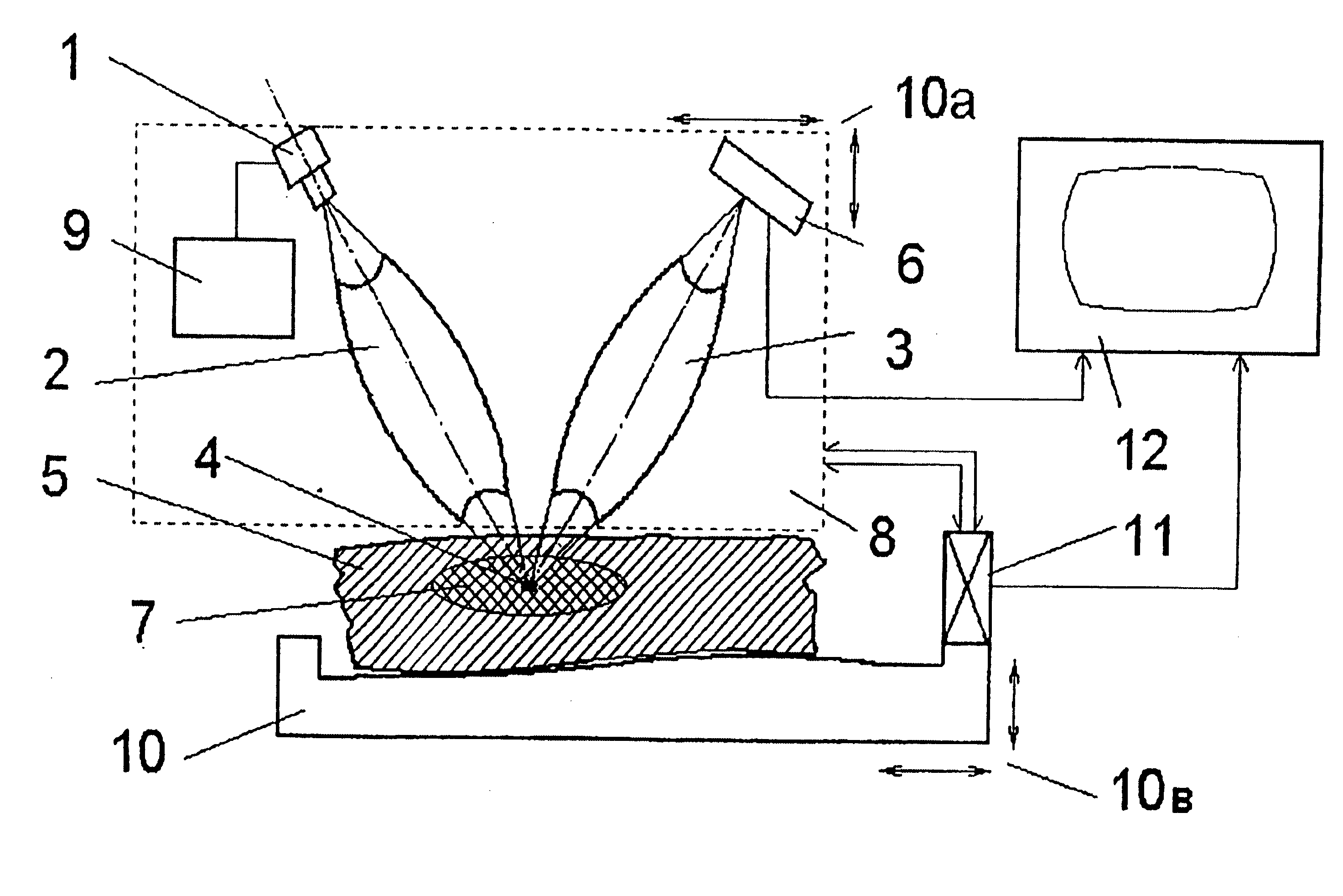
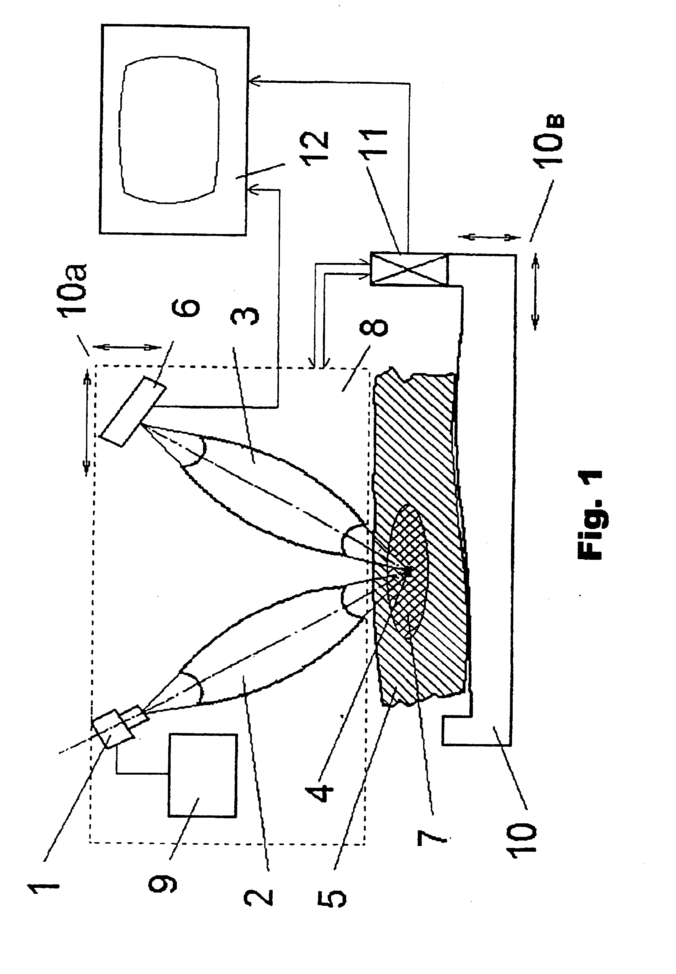
[0064] The methods proposed are accomplished using the device proposed in a following way.
[0065] Divergent X-ray radiation from quasi-point source 1, generating radiation with energy correspond...
PUM
 Abstract
Description
Claims
Application Information