
Methods and devices for cervix measurement
a technology for uterine cervix and measurement method, which is applied in the field of measuring the dimensions of the uterine cervix, can solve the problems of increasing the risk of infection to the mother and the fetus, discomfort for patients, and the method known as digital probing
- Summary
- Abstract
- Description
- Claims
- Application Information
AI Technical Summary
Benefits of technology
Problems solved by technology
Method used
Image

Examples
Embodiment Construction
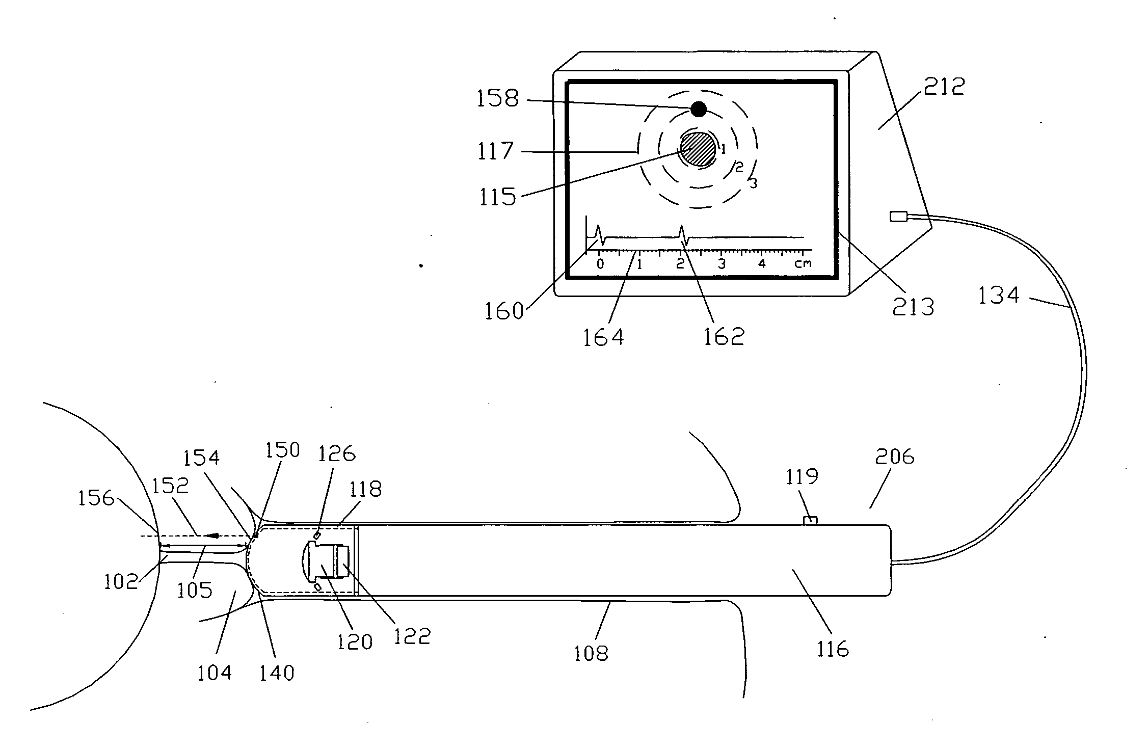
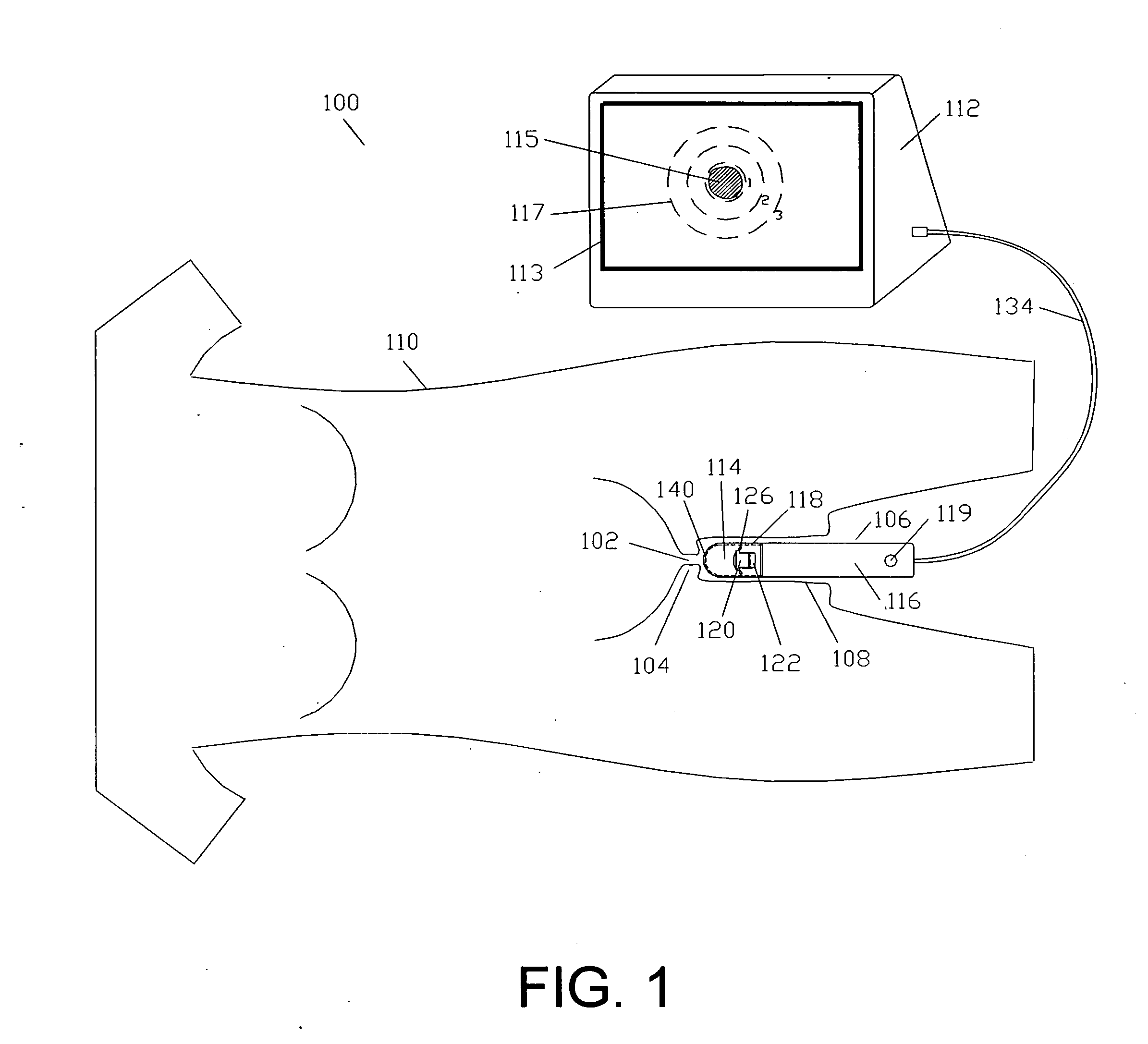
[0009] Referring to FIG. 1, the present invention includes a system 100 for measuring the diameter of the opening 102 of the cervix 104 in a manner that is non-invasive to the cervix. The system includes a probe 106 that is inserted into a vagina 108 of the patient 110 to gather data relating to the cervix 104, in particular, the diameter of the opening 102, and a monitoring unit 112 that is in communication with the probe 106 and receives image data from the probe 106 which is displayed to a health care provider located proximately to the patient. Accordingly, the patient need not have her cervix digitally probed.
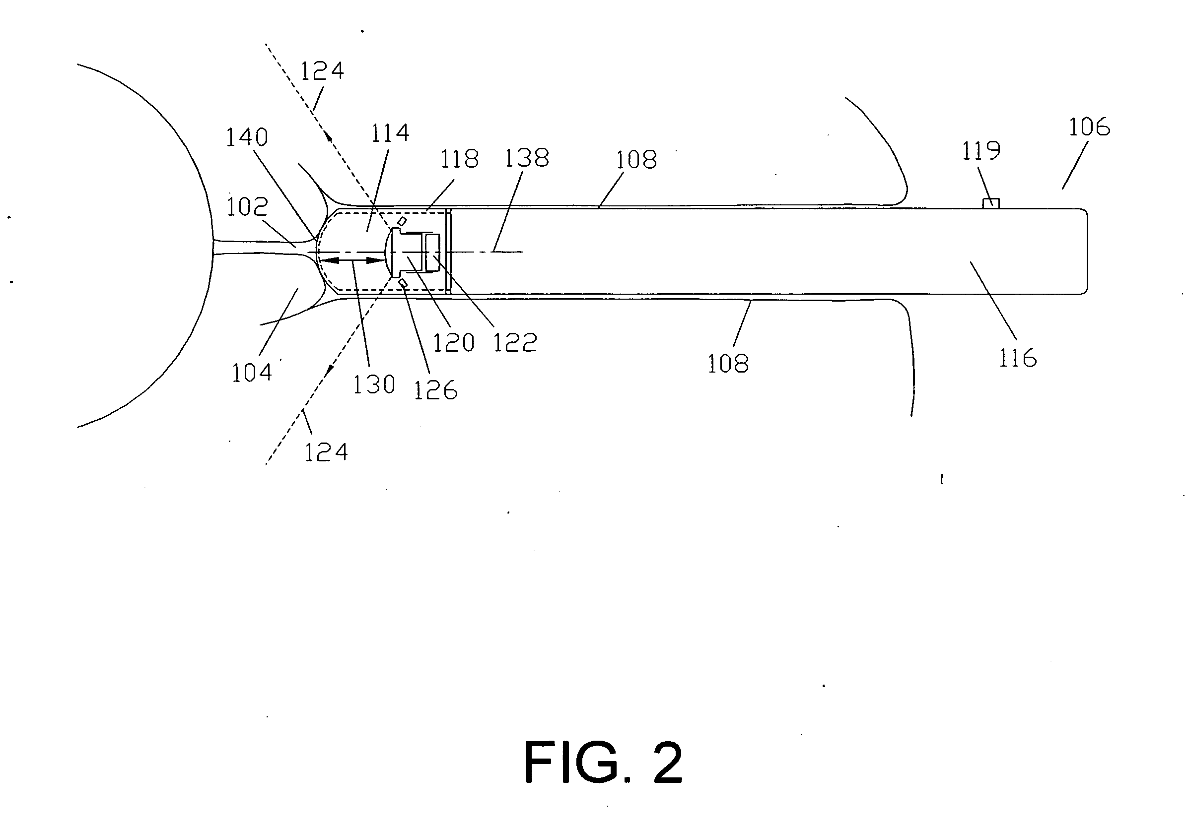
[0010] Referring to FIGS. 1 and 2, a preferred embodiment of the cervix probe 106 includes generally a distal probe head 114 and a proximal probe handle 116 extending proximally from the probe head. The probe head 114 includes a housing 118 encapsulating a lens 120 and a camera 122 that is proximal of the lens and adapted to capture image data of objects within a predeter...
PUM
 Abstract
Description
Claims
Application Information