Method and Apparatus for Inner Wall Extraction and Stent Strut Detection Using Intravascular Optical Coherence Tomography Imaging

a technology inner wall extraction, which is applied in the field of optical coherence tomography imaging techniques, can solve the problems of affecting the magnitude of nih after stent implantation, requiring a significant time investment, and manual processing was not always accurate, so as to achieve automatic and accurate determination

Inactive Publication Date: 2007-07-19
SIEMENS MEDICAL SOLUTIONS USA INC
View PDF2 Cites 162 Cited by
  • Summary
  • Abstract
  • Description
  • Claims
  • Application Information

AI Technical Summary

Benefits of technology

[0009] The present inventors have invented a method and apparatus for automatically detecting stent struts in an image obtained, for example, via OCT imaging techniques whereby the inner boundary of the artery wall is first detected automatically. One skilled in the art will recognize that the inner boundary of such an artery is also referred to as the lumen boundary of the artery. As such, the term lumen boundary will be used interchangeably herein to refer to the inner surface or wall of an artery. In one embodiment, detection of the lumen boundary may be accomplished, for example, by evolving a geometric shape, such as an ellipse, using either a region-based algorithm technique, a geodesic boundary-based algorithm tec

Problems solved by technology

More particularly, if the strut distribution is nonuniform, the resulting nonuniform distribution of the respective drug within the artery may also af

Method used


Image

  • Method and Apparatus for Inner Wall Extraction and Stent Strut Detection Using Intravascular Optical Coherence Tomography Imaging
  • Method and Apparatus for Inner Wall Extraction and Stent Strut Detection Using Intravascular Optical Coherence Tomography Imaging
  • Method and Apparatus for Inner Wall Extraction and Stent Strut Detection Using Intravascular Optical Coherence Tomography Imaging

Examples


Embodiment Construction

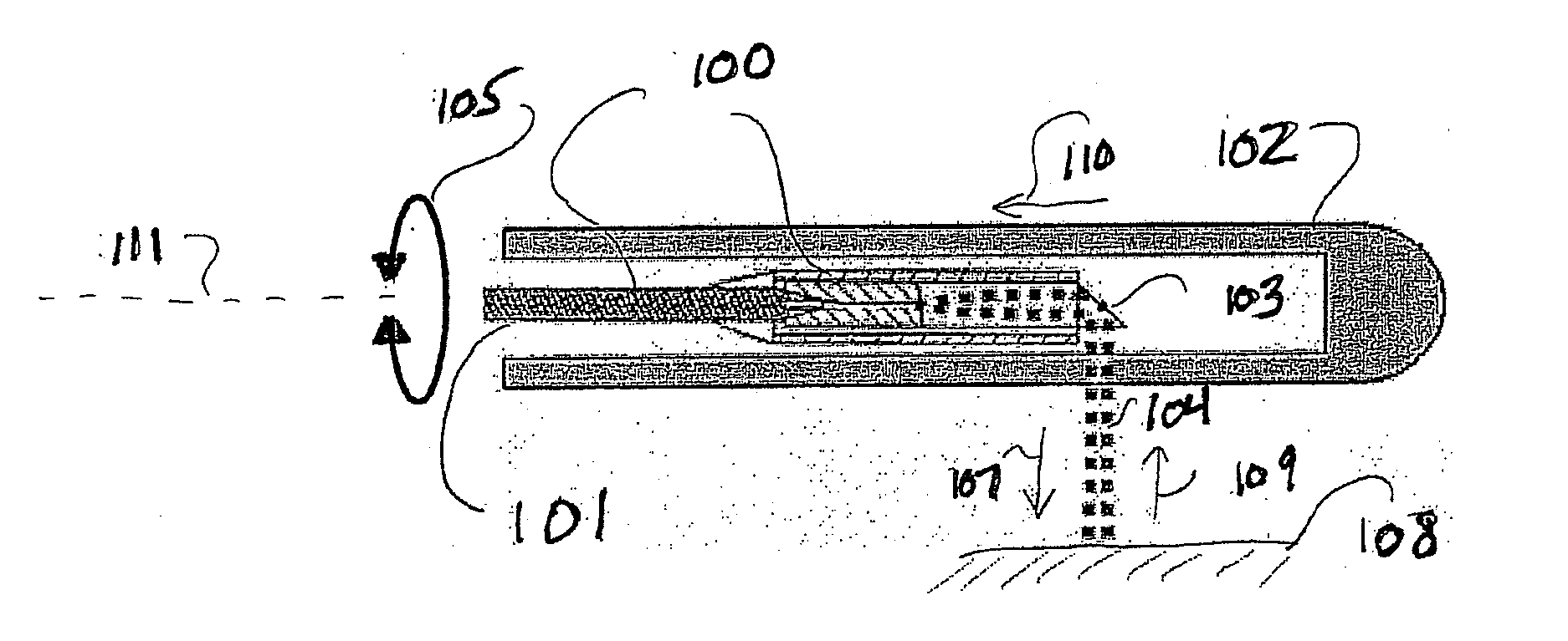
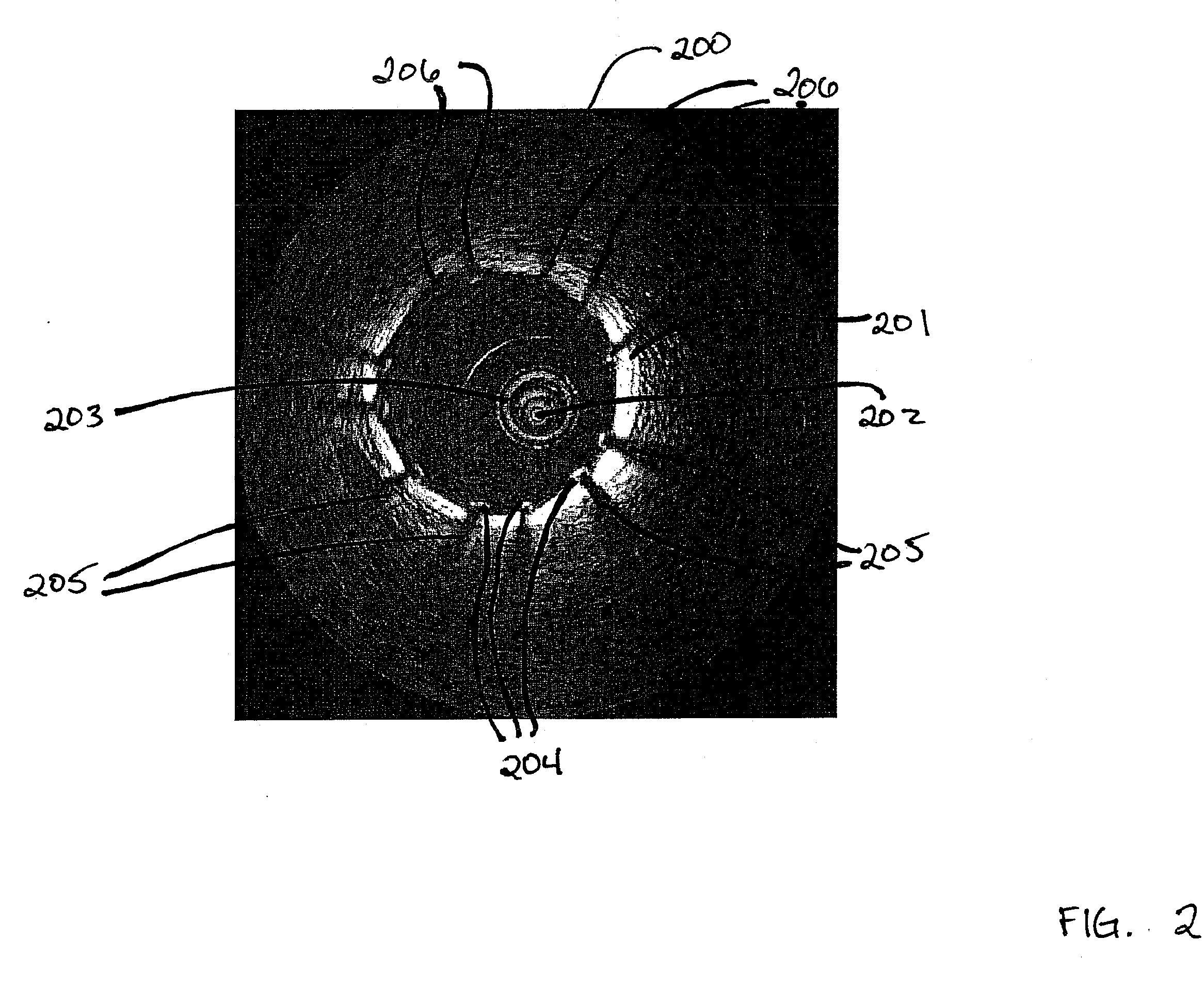
[0020]FIG. 2 shows a cross section image 200 showing an artery 201, which is for example a coronary artery in which a stent has been inserted. Image 200 is illustratively an image obtained via OCT scanning using optical or infrared light transmitted by sensor 202 that has been inserted into artery 201 using catheter 203. As one skilled in the art will recognize, the relatively bright portion of the image represents the wall of artery 201. FIG. 2 also shows cross section images of the struts 204 of a stent that has been inserted into the artery. Shadows, such as shadows 205, result when the stent blocks the optical or infrared signal originating from sensor 202. As discussed above, it is desirable to be able to locate the struts 204 in the image accurately but prior manual methods for identifying such struts were not accurate and typically took extensive time to perform. Accordingly, the present inventors have invented a method and apparatus for automatically identifying the struts i...


PUM


Abstract

A method and apparatus for automatically detecting stent struts in an image is disclosed whereby the inner boundary, or lumen, of an artery wall is first detected automatically and intensity profiles along rays in the image are determined. In one embodiment, detection of the lumen boundary may be accomplished, for example, by evolving a geometric shape, such as an ellipse, using a region-based algorithm technique, a geodesic boundary-based algorithm technique or a combination of the two techniques. Once the lumen boundary has been determined, in another embodiment, the stent struts are detected using a ray shooting algorithm whereby a ray is projected outward in the OCT image starting from the position in the image of the OCT sensor. The intensities of the pixels along the ray are used to detect the presence of a stent strut in the image.

Description

[0001] This patent application claims the benefit of U.S. Provisional Application No. 60 / 740,367, filed Nov. 29, 2005, which is hereby incorporated by reference herein in its entirety.BACKGROUND OF THE INVENTION [0002] The present invention relates generally to optical coherence tomography imaging techniques and, more particularly, to stent strut detection using such techniques. [0003] Coronary artery diseases, such as atherosclerosis, are a leading cause of death in the industrialized world. In particular, atherosclerotic plaques may cause narrowing or blockage of the coronary arteries, resulting in reduced blood supply to the heart tissue, which may sometimes lead to serious results such as heart attacks. Medical imaging techniques have greatly assisted the diagnosis and treatment of such coronary artery diseases. For example, coronary X-ray angiography, computed tomography angiography (CTA), magnetic resonance angiography (MRA), intravascular ultrasound (IVUS), and optical cohere...

Claims


Application Information

Patent Timeline
IPC IPC(8): A61B5/05
CPCA61B5/0066A61B5/6852A61B5/0086
Inventor UNAL, GOZDEYANG, YANSLABAUGH, GREGORY G.FANG, TONG
Owner SIEMENS MEDICAL SOLUTIONS USA INC
Why Eureka
Try Eureka