Method for ascertaining the position of a medical instrument in a body

a technology for medical instruments and head positions, applied in medical science, surgical navigation systems, sensors, etc., can solve the problems of radiation load on the patient and the physician, and achieve the effect of not having to increase the size of the medical instrumen

Inactive Publication Date: 2009-12-31
BRAINLAB
View PDF31 Cites 3 Cited by
  • Summary
  • Abstract
  • Description
  • Claims
  • Application Information

AI Technical Summary

Benefits of technology

[0006]It is therefore the object of the present invention to provide a method and device for ascertaining the position of the head of a medical instrument in a body, in which a radiation load can be reduced and in which in particular the overall size of the medical instrument does not have to be increased. The latter enables a broader range of application for the instrument.

Problems solved by technology

However, this procedure incurs a radiation load both for the patient and the physician.
This can necessitate a rather large instrument.

Method used


Image

  • Method for ascertaining the position of a medical instrument in a body
  • Method for ascertaining the position of a medical instrument in a body
  • Method for ascertaining the position of a medical instrument in a body

Examples


Embodiment Construction

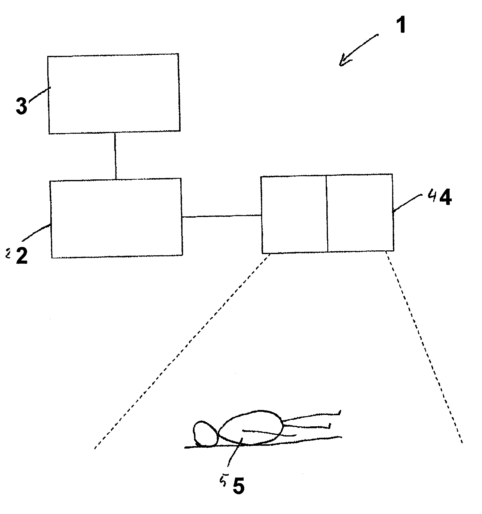
[0037]FIG. 1 schematically shows an operation system 1 comprising a computational unit 2, a monitor 3 and a 3D infrared camera 4. A patient 5 to be examined is situated within the detection range of the camera 4 shown by broken lines.

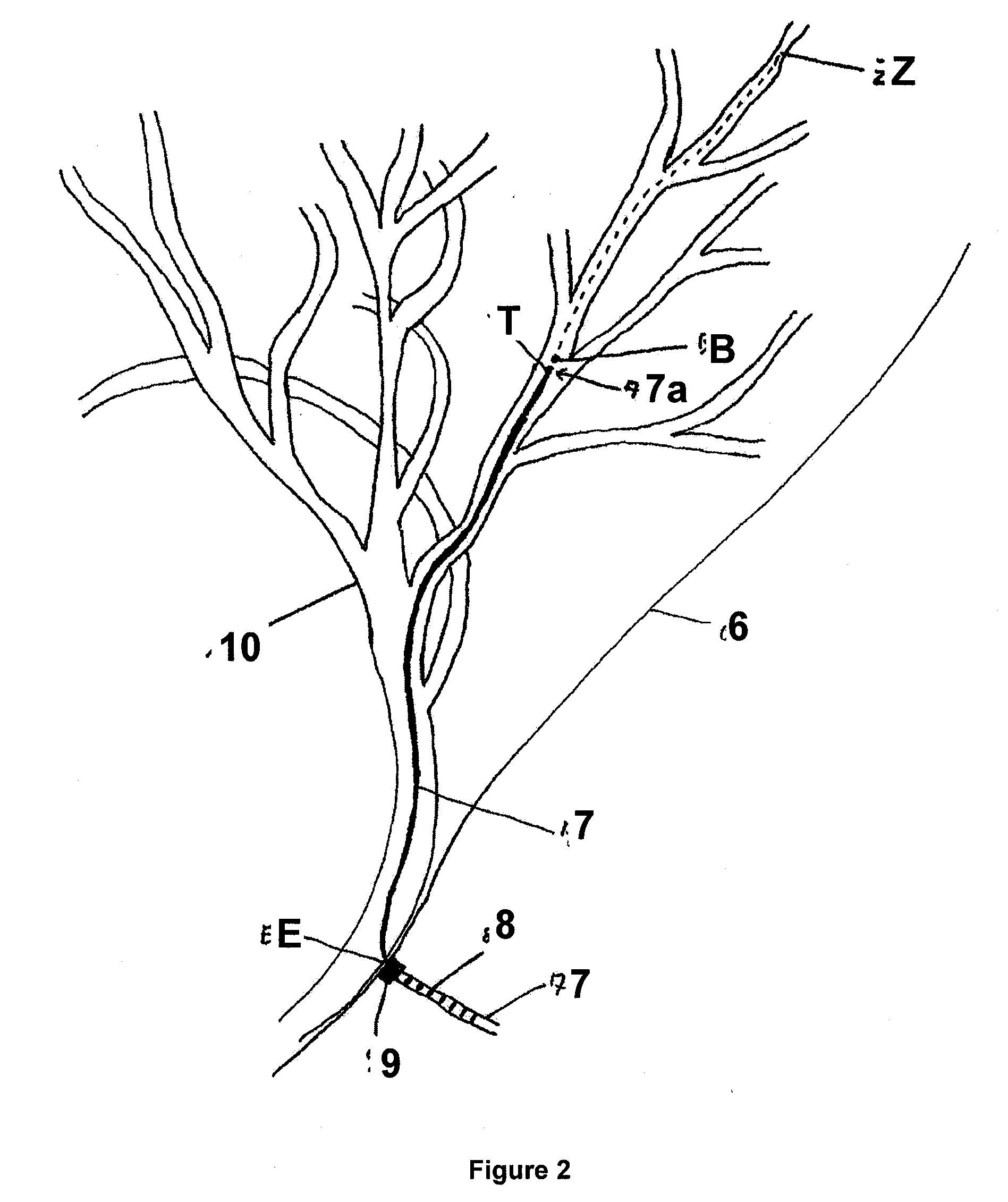
[0038]FIG. 2 shows a part of the vascular system 10 of the patient 5 which is situated below the skin 6. The vascular system 10 is to be examined at a position Z by means of an endoscope 7. The endoscope 7 is inserted into the vascular system 10, which lies directly below the skin 6, at an entry point E. The structure, i.e. the three-dimensional configuration, of the vascular system 10 has been ascertained, by means of a computer tomograph, before the endoscope 7 is inserted. The planned path between the entry point E and the target point Z is shown by a broken line.

[0039]A regular marking 8 is situated on the endoscope 7, in the form of equidistant bars which are detected by a measuring device 9 at the entry point E. Two light sources and two photodete...


PUM


Abstract

The present invention relates to a method for ascertaining the position of the head of a medical instrument in a vascular system, wherein the instrument can be inserted into the vascular system of a body and describes a path at least partially inside the vascular system, and the position of the instrument head is calculated from structural data, length data and the relative position between a reference point and the vascular system, wherein the instrument is guided past the reference point, the structural data represents the structure of the vascular system, and the length data represents the length of the path between the reference point and the instrument head.

Description

RELATED APPLICATION DATA[0001]This application claims the priority of U.S. Provisional Application No. 61 / 094,894, filed on Sep. 6, 2008, which is hereby incorporated in its entirety by reference.FIELD OF THE INVENTION[0002]The invention relates to: a method for ascertaining the position of a head of a medical instrument in a body, in particular in a vascular system; a program for performing the method; a storage medium on which the program is stored; a device for ascertaining the position of the head of a medical instrument in a body; and an operation system comprising a medical instrument and such a device.BACKGROUND OF THE INVENTION[0003]In an operation or examination, medical instruments are often used which are inserted into the body to be operated on, in particular into a vascular system, wherein it is important to know the position of the head, i.e. the inserted end, of the medical instrument.[0004]In previous practice, the position has been determined using real-time imaging...

Claims


Application Information

Patent Timeline
Patent Type & Authority Applications(United States)
IPC IPC(8): A61B5/05
CPCA61B5/06A61B5/064A61B6/12A61B2019/5289A61B19/5244A61B2019/461A61B19/5212A61B34/20A61B2090/364A61B90/361A61B2090/061
Inventor SCHMIDT, ROBERTURBAN, ALEXANDERVOLLMER, FRITZ
Owner BRAINLAB
Why Eureka
Try Eureka