
Endocardial mapping system
a mapping system and endocardial technology, applied in the field of endocardial mapping system, can solve the problems of inability to accurately resolve the location of ectopic tissue masses, the two-dimensional mapping of electrical potentials at the endocardial surface generated by these traditional processes suffer many defects, and the resolution of traditional systems is limited
- Summary
- Abstract
- Description
- Claims
- Application Information
AI Technical Summary
Benefits of technology
Problems solved by technology
Method used
Image

Examples
Embodiment Construction
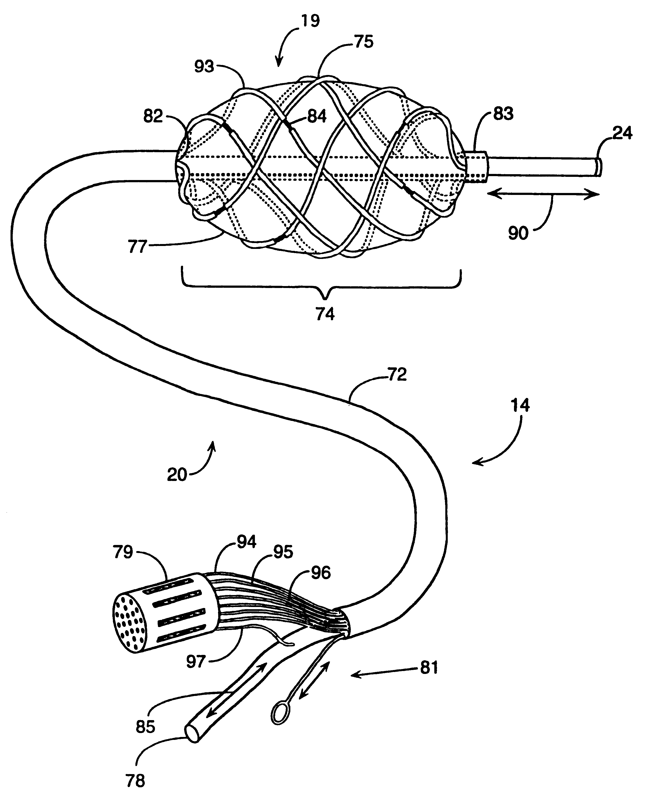
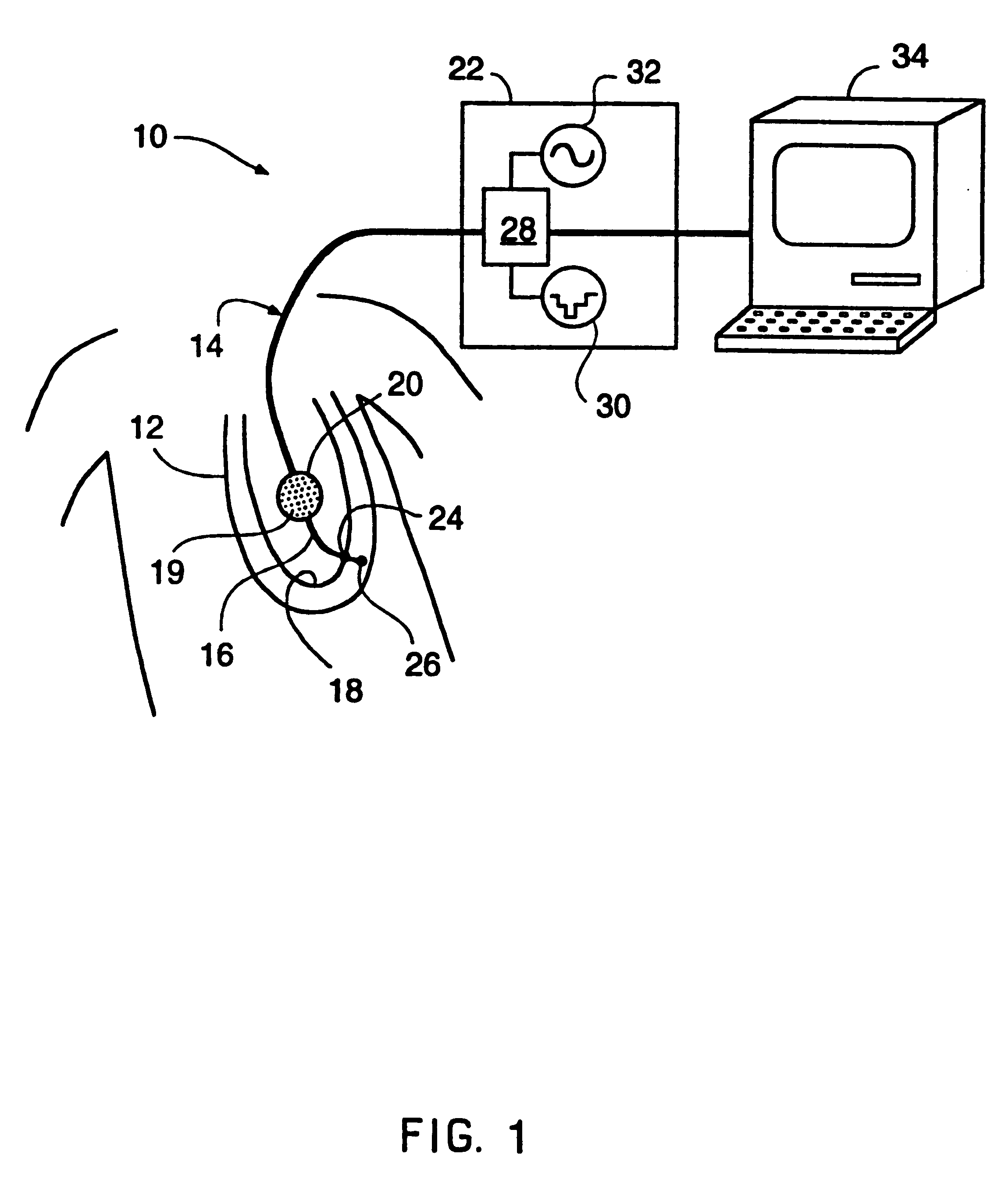
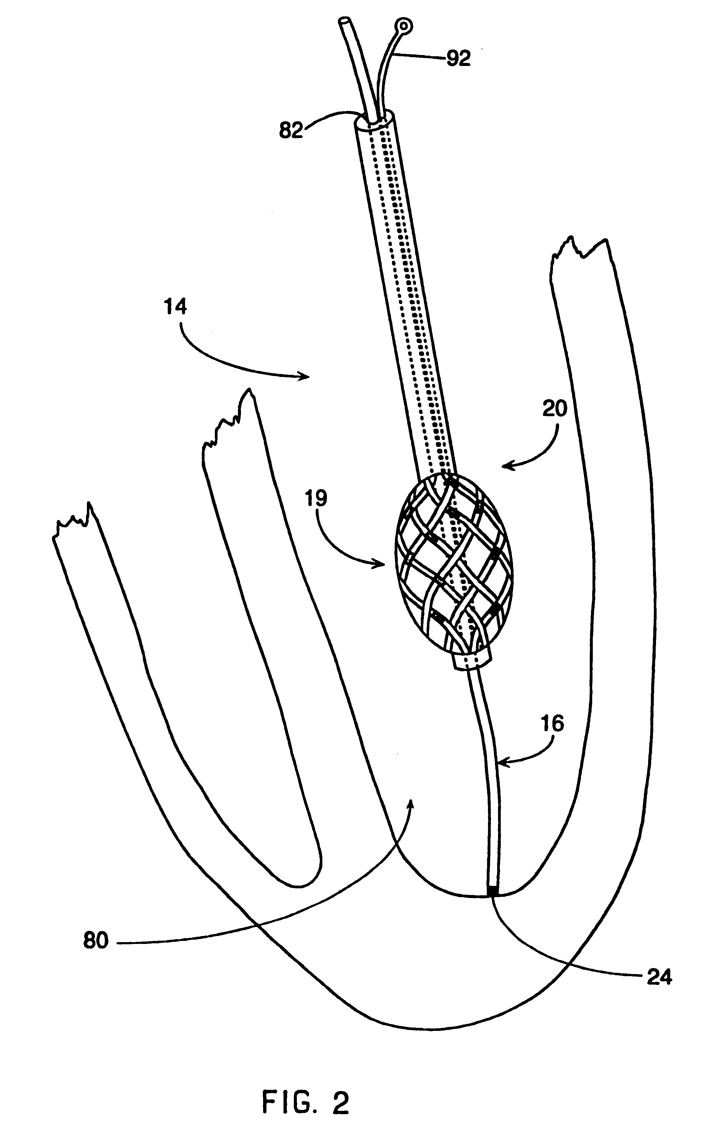
In general, the system of the present invention is used for mapping the electrical activity of the interior surface of a heart chamber 80. The mapping catheter assembly 14 includes a flexible lead body 72 connected to a deformable distal lead body 74. The deformable distal lead body 74 can be formed into a stable space filling geometric shape after introduction into the heart cavity 80. This deformable distal lead body 74 includes an electrode array 19 defining a number of electrode sites. The mapping catheter assembly 14 also includes a reference electrode preferably placed on a reference catheter 16 which passes through a central lumen 82 formed in the flexible lead body 72 and the distal lead body 74. The reference catheter assembly 16 has a distal tip electrode assembly 24 which may be used to probe the heart wall. This distal contact electrode assembly 24 provides a surface electrical reference for calibration. The physical length of the reference catheter 16 taken with the pos...
PUM
 Abstract
Description
Claims
Application Information