Biopsy device with replaceable probe incorporating static vacuum source dual valve sample stacking retrieval and saline flush

a biopsy device and vacuum source technology, applied in the field of biopsy devices, can solve the problems of high cost and high level of trauma to the patient, open biopsy carries a relatively higher risk of infection and bleeding, and is difficult to read future mammograms

Inactive Publication Date: 2010-02-16
DEVICOR MEDICAL PROD
View PDF29 Cites 59 Cited by
  • Summary
  • Abstract
  • Description
  • Claims
  • Application Information

AI Technical Summary

Benefits of technology

[0014]The present invention addresses these and other problems of the prior art by providing a biopsy device that has a needle with a probe tube defining a cutter lumen, a sample aperture formed in the probe tube, a barrier defining a first fluid passage and a second fluid passage that both distally-terminate at the sample aperture. A motorized mechanism axially translates a cutter tube within the probe tube across the sample aperture to sever tissue prolapsed therein to axially translate the cutter tube. One of the first and second fluid passages is defined within the cutter tube and the other is defined between an outer surface of the cutter tube and an inner surface of the probe tube. Advantageously, a flush valve assembly responds to a flush control and to the distally positioned cutter tube to couple either the first or second fluid passage to a fluid supply while the other is at a lower pressure so that the needle is flushed. Thereby, tissue debris or coagulated blood may be flushed so that repeated tissue samples may be taken without impediment. However, the saline flush is selectively employed at the user's discretion, providing an economical reduction in the usage of saline and a corresponding reduction in the overall size of the fluid collection reservoir. It is also believed that certain pathology analyses would benefit from not subjecting tissue samples to a saline flush.
[0015]In another aspect of the invention, a core biopsy device has a probe assembly with a probe support structure that holds a probe having a side aperture. A cutter tube is slidingly received by the probe and sized to translate across the side aperture to sever prolapsed tissue. A hand piece includes a hand piece support structure having a lateral engaging portion that receives the probe assembly. Thereby, an economical incorporation of a replaceable probe and cutter tube into a laterally mounted assembly allows reuse of a powered hand piece, yet also provides an advantageous saline flush capability of the probe assembly.

Problems solved by technology

An open biopsy is a surgical procedure that is usually done as an outpatient procedure in a hospital or a surgical center, involving both high cost and a high level of trauma to the patient.
Open biopsy carries a relatively higher risk of infection and bleeding than does percutaneous biopsy, and the disfigurement that sometimes results from an open biopsy may make it difficult to read future mammograms.
Further, the aesthetic considerations of the patient make open biopsy even less appealing due to the risk of disfigurement.
Given that a high percentage of biopsies show that the suspicious tissue mass is not cancerous, the downsides of the open biopsy procedure render this method inappropriate in many cases.
However, a shortcoming of FNA is that only a small number of cells are obtained through the procedure, rendering it relatively less useful in analyzing the suspicious tissue and making an assessment of the progression of the cancer less simple if the sample is found to be malignant.
With a long probe, the rate of sample taking is limited not only by the time required to rotate or reposition the probe but also by the time needed to translate the cutter.
Thus, the surgeon is challenged to maintain the biopsy probe at a desired surgical site while manipulating the patient's breast.
Frequently, a surgeon may encounter an area of dense tissue that is more difficult to penetrate than the surrounding tissue during core needle biopsy.
In particular, the lesion or tissue mass being targeted in the biopsy procedure may be difficult to penetrate, requiring the physician to push the biopsy needle with considerable force and / or speed in an attempt to penetrate the lesion and collect a sample.
However, due to the length of the firing stroke of such devices, which may be as long as 0.75 inches, it is nearly impossible for the surgeon to control the travel of the needle after firing.
Consequently, the long needle stroke may cause uncertainty as to the needle tip location post fire.
This may cause the surgeon to obtain a sample from the wrong area.
In addition to missing the targeted tissue, long firing strokes may cause the needle to puncture the chest wall or pierce the skin, particularly when the targeted area is near the patient's chest wall.
Even if the skin is not pierced, the long travel of the needle, along with the likelihood that the needle will be pushed off course by the force of the firing stroke, may lead to needlessly increased trauma for the patient.
These spring-fired biopsy devices also yield a single sample per insertion, thus limiting the amount of diagnostic and therapeutic treatment that may be achieved without the increased discomfort and tissue trauma from repeated insertions.

Method used


Image

  • Biopsy device with replaceable probe incorporating static vacuum source dual valve sample stacking retrieval and saline flush
  • Biopsy device with replaceable probe incorporating static vacuum source dual valve sample stacking retrieval and saline flush
  • Biopsy device with replaceable probe incorporating static vacuum source dual valve sample stacking retrieval and saline flush

Examples


Embodiment Construction

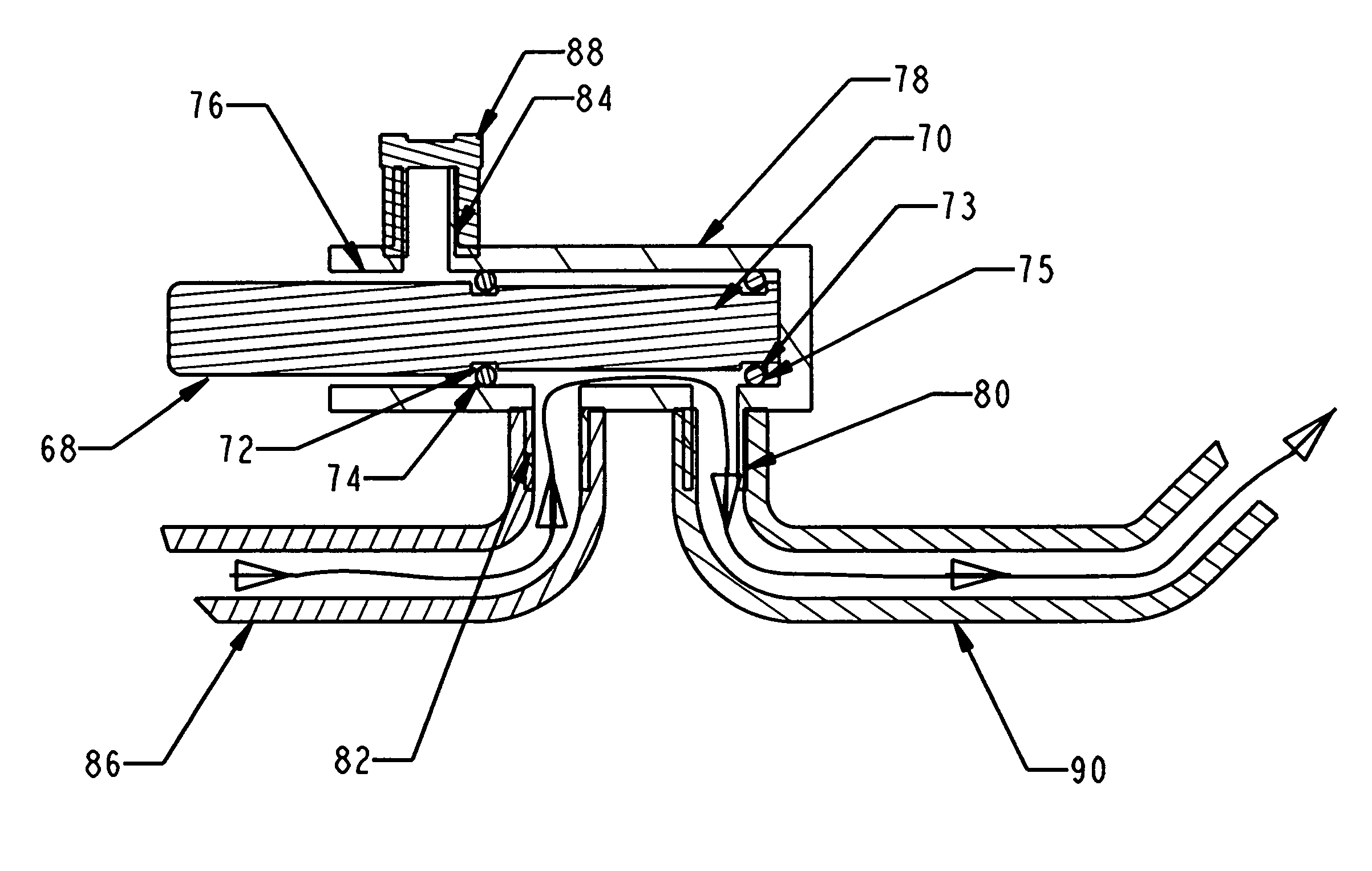
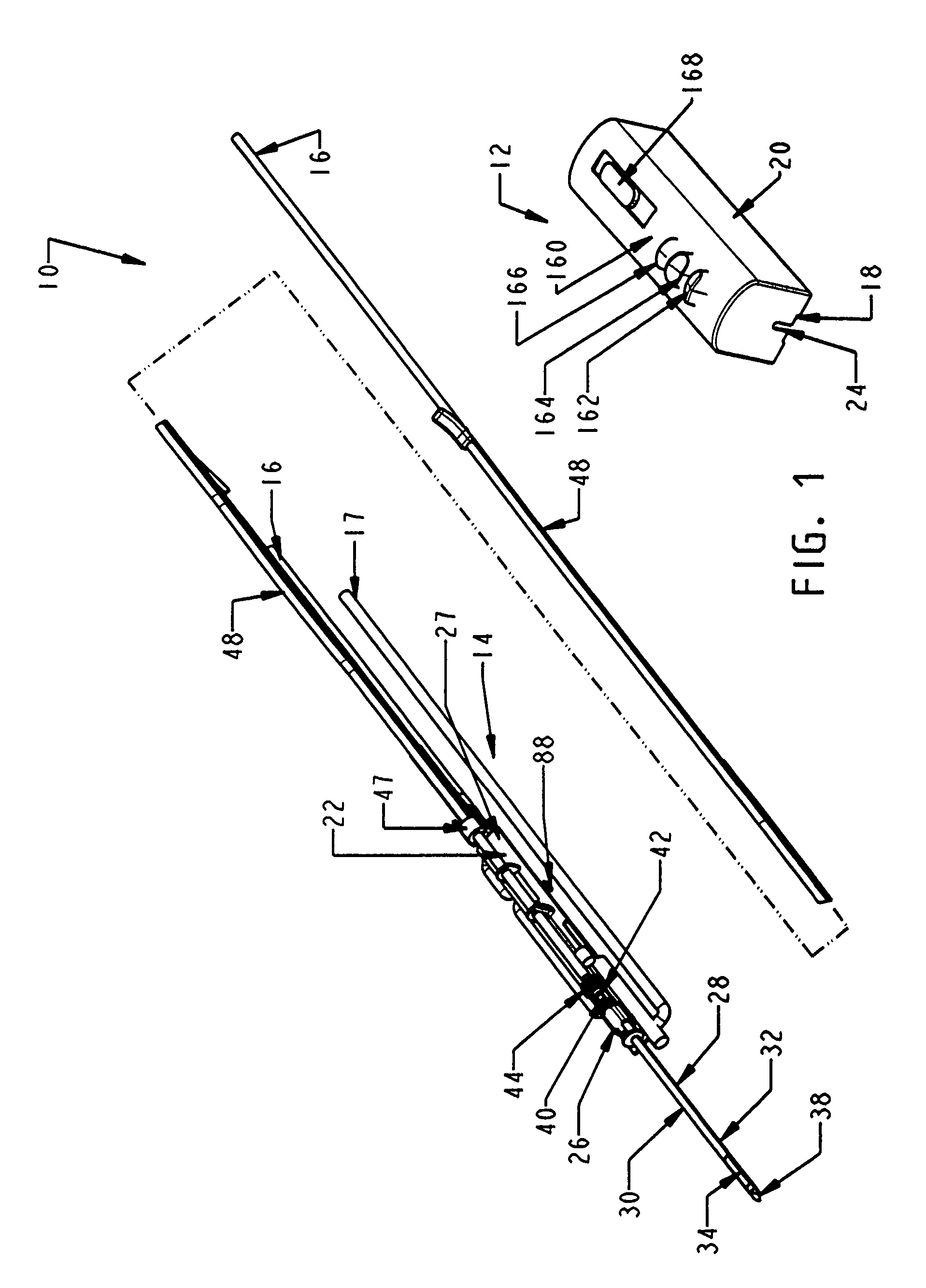
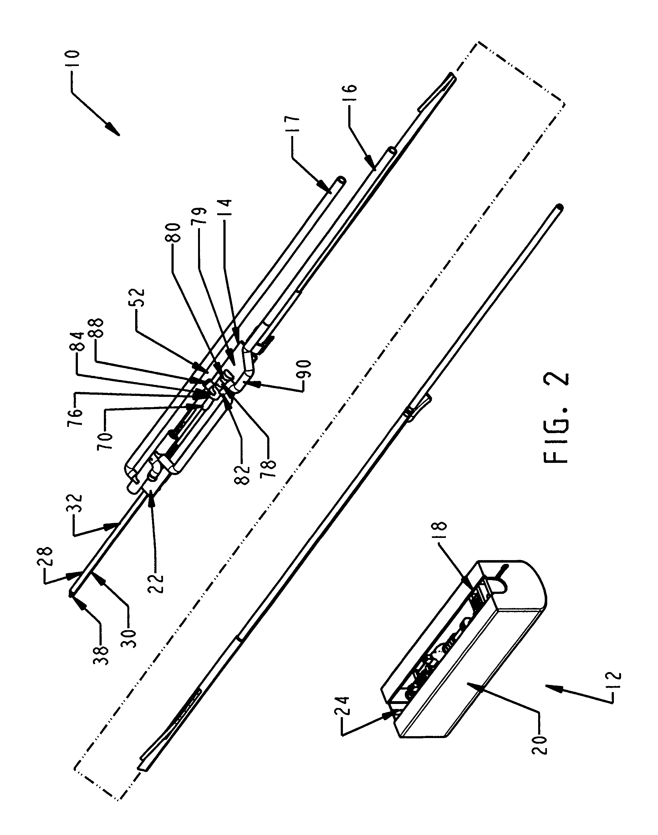
[0042]In FIGS. 1-2, a biopsy device 10 has a reusable hand piece 12 and a disposable probe 14 that enables economical taking of multiple percutaneous core biopsy samples by accessing a standard medical vacuum pump or wall-mounted vacuum access port (not shown) through an interfacing vacuum conduit 16. In addition, the biopsy device 10 advantageously incorporates a saline flush capability received from saline supply conduit 17. In the illustrative version, the reusable hand piece 12 is self-powered and suitable for use in conjunction with ultrasonic diagnostic imaging. The disposable probe 14 reduces the portion of biopsy device 10 that requires protective packaging to avoid contact with sharp surfaces and to keep it sterile prior to use. Further economy is accomplished by reducing the portion of the biopsy device 10 that is disposed as medical waste between uses. Movable components of the disposable probe 14 are advantageously locked until mounted in an access trough 18 formed in a ...


PUM


Abstract

A biopsy device and method are provided for obtaining a tissue sample, such as a breast tissue biopsy sample. The biopsy device includes a disposable probe assembly with an outer cannula having a distal piercing tip, a cutter lumen, and a cutter tube that rotates and translates past a side aperture in the outer cannula to sever a tissue sample. The biopsy device also includes a reusable hand piece with an integral motor and power source to make a convenient, untethered control for use with ultrasonic imaging. The reusable hand piece incorporates a probe oscillation mode to assist when inserting the distal piercing tip into tissue. A saline valve positioned by the reusable hand piece communicates a saline supply through the probe assembly to perform saline flush of the cutter tube and outer cannula.

Description

CROSS REFERENCE TO RELATED APPLICATIONS[0001]The present application is related to the co-pending and commonly-owned U.S. patent application Ser. No. 11 / 198,558 “BIOPSY DEVICE WITH REPLACEABLE PROBE AND INCORPORATING VIBRATION INSERTION ASSIST AND STATIC VACUUM SOURCE SAMPLE STACKING RETRIEVAL” to Hibner et al., filed 8 Aug. 2005, the disclosure of which is hereby incorporated by reference in its entirety.FIELD OF THE INVENTION[0002]The present invention relates in general to biopsy devices, and more particularly to biopsy devices having a cutter for severing tissue, and even more particularly to biopsy devices for multiple sampling with a probe remaining inserted.BACKGROUND OF THE INVENTION[0003]When a suspicious tissue mass is discovered in a patient's breast through examination, ultrasound, MRI, X-ray imaging or the like, it is often necessary to perform a biopsy procedure to remove one or more samples of that tissue in order to determine whether the mass contains cancerous cells...

Claims


Application Information

Patent Timeline
Patent Type & Authority Patents(United States)
IPC IPC(8): A61B10/00
CPCA61B10/0275A61B10/0283A61B10/04A61B2010/0208A61B2010/0225A61B2217/007A61B2017/0023A61B2017/00398A61B2017/00734A61B2217/005A61B2017/0046
Inventor HIBNER, JOHN A.
Owner DEVICOR MEDICAL PROD
Why Eureka
Try Eureka