Method for panoramic imaging ophthalmology protomerite detected by ultrasound biological microscopes

A panoramic imaging and microscope technology, applied in fundus mirror, image enhancement, image analysis, etc., can solve the problems of sparse features, large rotation angle, large noise, etc., and achieve the effect of small geometric distortion, fast running speed and high resolution

Active Publication Date: 2008-10-22
TIANJIN SUOWEI ELECTRONICS TECH
View PDF0 Cites 16 Cited by
  • Summary
  • Abstract
  • Description
  • Claims
  • Application Information

AI Technical Summary

Problems solved by technology

However, for the images of the anterior segment of the eyeball, due to its sparse features, large noise, large deformation between adjacent images, and relatively large rotation angle, the general image stitching method cannot be applied.

Method used


Image

  • Method for panoramic imaging ophthalmology protomerite detected by ultrasound biological microscopes
  • Method for panoramic imaging ophthalmology protomerite detected by ultrasound biological microscopes
  • Method for panoramic imaging ophthalmology protomerite detected by ultrasound biological microscopes

Examples


Embodiment Construction

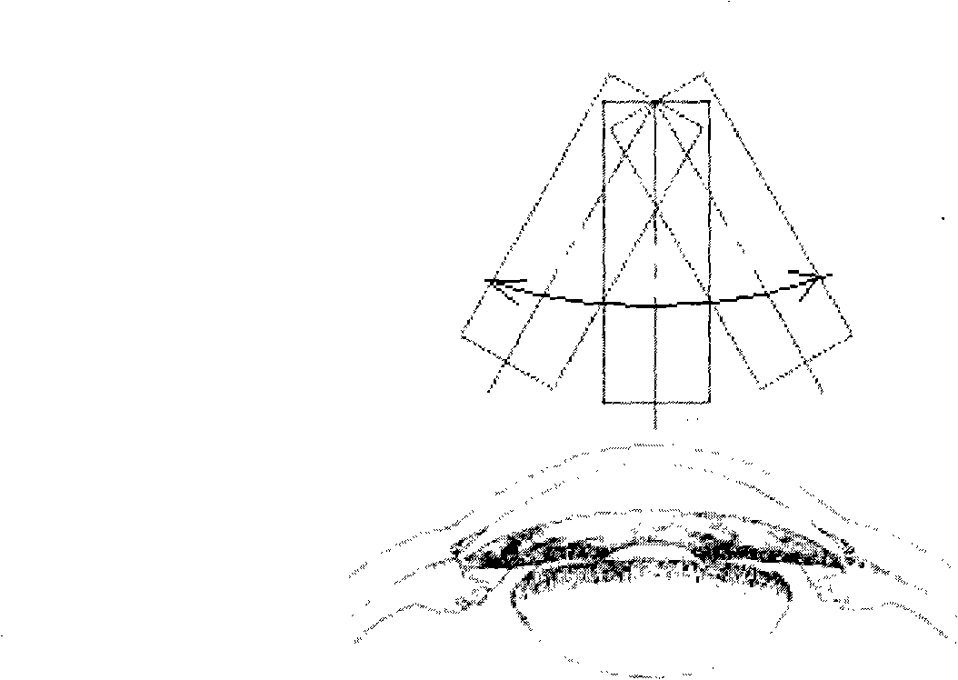
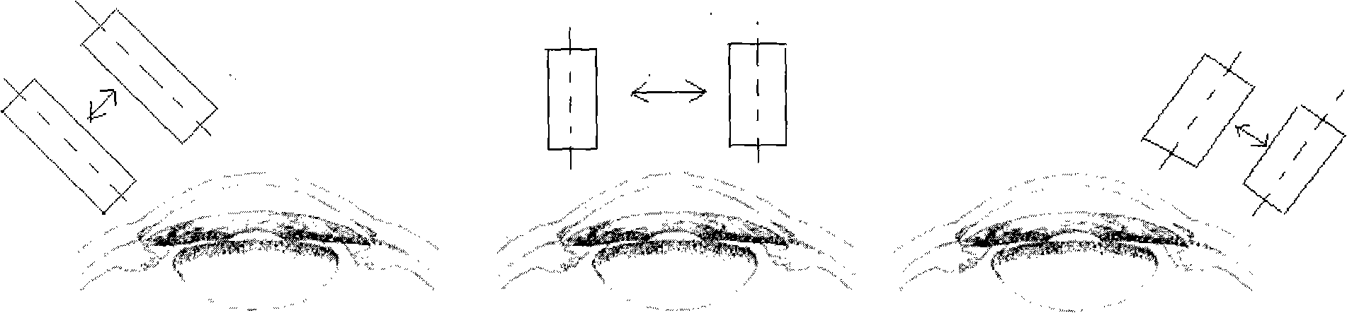
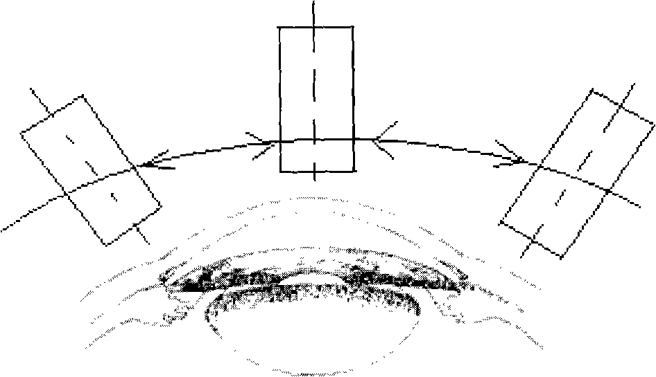
[0047] The present invention uses the image registration and splicing method based on the image features of the anterior segment to synthesize a complete anterior segment section image from overlapping images on the same meridian plane of the eyeball. figure 2 In the scanning method shown, a group of continuous images of the anterior segment are obtained along a certain meridian of the cornea by using a linear scanning ultrasonic biomicroscope. The number of images is four, and the scanning results are as follows: Figure 7 As shown, they are pictures a, b, c, and d respectively, pictures a and b, b and c, c and d each have overlapping parts, and the size of each picture is 512×512 pixels, which is an 8-bit grayscale image , the brightness of the image ranges from 0 to 255.

[0048] then to Figure 7 The four previous images are filtered according to the image mean value, and the filter window size is 9×9, then the filtered image is:

[0049] f ...


PUM


Abstract

The invention discloses a panoramic view imaging method for realizing ophthalmonogy prosthomere by UBM. Firstly, at least four continuous pictures with two adjacent pictures mutually overlapped are obtained along the cornea longitude direction by linear UBM; then a whole eyeball prosthomere picture is spliced with the pictures by image filtering, binarization, lines clustering, joint location searching and image fusion. The method has the image precision of linear UBM, at the same time, the eyeball prosthomere panoramic view imaging is realized.

Description

technical field [0001] The invention relates to a panoramic imaging method for anterior segment examination in ophthalmology, in particular to a method for realizing panoramic imaging of anterior segment in ophthalmology on the basis of linear UBM examination through image registration and splicing based on anterior segment features. Background technique [0002] Ultrasonic biomicroscopy (UBM) currently used for anterior segment examination in ophthalmology all uses mechanical scanning, which is mainly divided into sector scanning (see figure 1 ), linear scan (see figure 2 ) and arc scan (see image 3 ) three categories. The sector scanning structure is the simplest, but during the scanning process, the working distance changes greatly, the resolution of the entire image varies greatly, and there is a certain geometric distortion. The arc scanning structure is the most complex, but during the scanning process, the working distance changes the least, so it can ensure that...

Claims


Application Information

Patent Timeline
Patent Type & Authority Applications(China)
IPC IPC(8): A61B3/13G06F19/00G06T7/00G06T3/40G06T5/50G06T7/33
Inventor 王宁利王雪乔王健发
Owner TIANJIN SUOWEI ELECTRONICS TECH
Why Eureka
Try Eureka