Three-dimensional segmentation method for intravascular ultrasound image sequence

An ultrasound image, vascular lumen technology, applied in the field of medical imaging, can solve the problems of low processing efficiency, poor repeatability, time-consuming, etc., and achieve the effect of improving processing efficiency and shortening processing time

Inactive Publication Date: 2011-02-02
NORTH CHINA ELECTRIC POWER UNIV (BAODING)
View PDF4 Cites 21 Cited by
  • Summary
  • Abstract
  • Description
  • Claims
  • Application Information

AI Technical Summary

Problems solved by technology

Since the IVUS image sequence often includes thousands of frames, this method is not only time-consuming, but also has poor repeatability, and the segmentation results largely depend on the operator's experience and subjective factors
The existing automatic segmentation methods using digital image processing technology are all serially processed in two-dimensional space, that is, each frame image in the IVUS sequence is segmented frame by frame, so the processing efficiency is low

Method used


Image

  • Three-dimensional segmentation method for intravascular ultrasound image sequence
  • Three-dimensional segmentation method for intravascular ultrasound image sequence
  • Three-dimensional segmentation method for intravascular ultrasound image sequence

Examples


Embodiment Construction

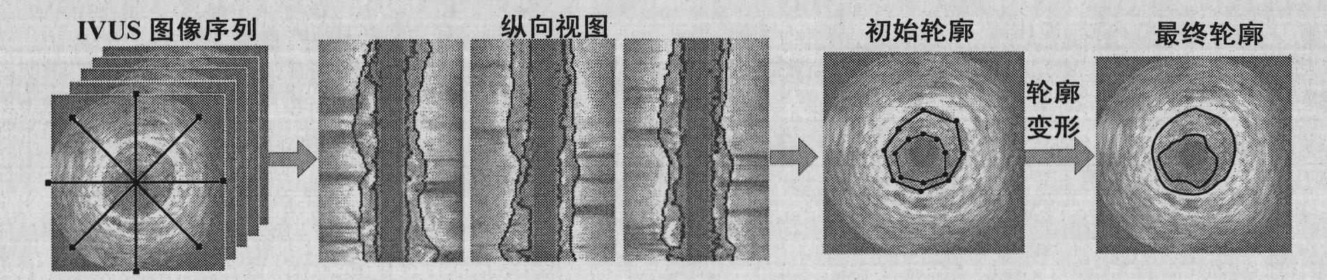
[0035] as attached figure 1 Shown, the step of the inventive method comprises:

[0036] (1) Preprocessing the original IVUS image, including filtering and denoising and removing halo artifacts:

[0037] Firstly, two common preprocessing methods, median filtering and Gaussian smoothing, are used to reduce salt and pepper noise and random noise in IVUS images.

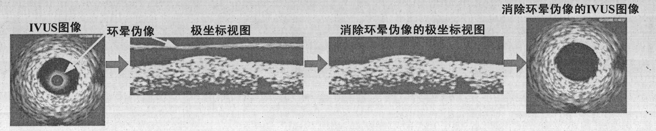
[0038] Then the polar coordinate transformation method is used to remove the halo artifact, the specific method is as follows:

[0039] First, perform polar coordinate transformation on each frame of IVUS image to obtain its polar coordinate view, as shown in the attached figure 2 shown. It can be seen that the halo artifact is fixedly located in the upper part of the polar coordinate view. Then, remove the halo artifact in the polar view according to the following formula:

[0040] I ′ ( r , θ ) = ...


PUM


Abstract

The invention discloses a three-dimensional segmentation method for an intravascular ultrasound (IVUS) image sequence, which is used for improving the segmentation processing efficiency of an image sequence. The technical scheme comprises the following steps of: first, performing preprocessing of filtering noise and inhibiting a ring halo pseudomorphism on an original image; then, acquiring four longitudinal views of the IVUS image sequence and extracting intravascular cavity boundaries and intermediate-outer film boundaries from the longitudinal views; next, acquiring initial boundaries in transverse views by mapping boundary curves to IVUS images of each frame; and finally, acquiring the intravascular cavity boundaries and the intermediate-outer film boundaries in the IVUS images of the each frame finally by maximizing an energy function and discontinuously deforming the initial boundaries. Compared with a conventional method, the three-dimensional segmentation method has the following advantages of: first, capacity of utilizing the information of an overall image sequence; and second, capacity of finishing segmenting images of the each frame at the same time and realizing parallel processing of the overall image sequence, so as to greatly improve processing efficiency and shorten processing time.

Description

technical field [0001] The invention relates to a three-dimensional segmentation method of an intravascular ultrasonic image sequence, which can realize parallel segmentation of an IVUS image sequence, and belongs to the technical field of medical imaging. Background technique [0002] Intravascular Ultrasound (IVUS) imaging technology can reflect changes in the lumen of blood vessels, as well as the cross-sectional structure of the lumen including plaques, the thickness and shape of blood vessel walls, and plaque components. The blood vessel wall presents a three-layer ring structure with different echoes on the IVUS image, and the front and rear frames in the image sequence are very similar and have a strong correlation. [0003] Segmenting IVUS images to extract the lumen boundary and media-adventitia boundary (including possible plaque edges) is an important step in quantitative analysis and three-dimensional reconstruction of blood vessels. The quality of segmentation d...

Claims


Application Information

Patent Timeline
Patent Type & Authority Applications(China)
IPC IPC(8): G06T5/00G06T17/00
Inventor 孙正杨宇
Owner NORTH CHINA ELECTRIC POWER UNIV (BAODING)
Why Eureka
Try Eureka