A Liver CT Atlas Segmentation Method and System Based on High Precision Registration

A high-precision, atlas technology, applied in the field of medical image processing, can solve the problem of large amount of calculation of 3D volume data

Active Publication Date: 2020-08-25
SOUTH CENTRAL UNIVERSITY FOR NATIONALITIES
View PDF8 Cites 0 Cited by
  • Summary
  • Abstract
  • Description
  • Claims
  • Application Information

AI Technical Summary

Problems solved by technology

Finally, it converges to the desired target contour, which is computationally expensive for 3D volume data

Method used


Image

  • A Liver CT Atlas Segmentation Method and System Based on High Precision Registration
  • A Liver CT Atlas Segmentation Method and System Based on High Precision Registration
  • A Liver CT Atlas Segmentation Method and System Based on High Precision Registration

Examples


Embodiment 1

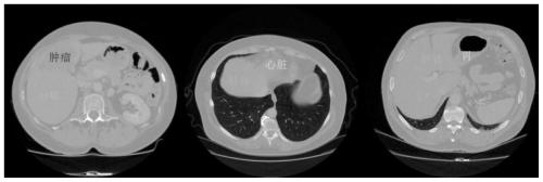
[0075] The embodiment of the present invention discloses a liver CT atlas segmentation method based on high-precision registration. The image data used in the embodiment of the present invention comes from liver CT scans of the SLIVER07 data set and the IRCAD data set. In order to ensure that the sample size of the atlas set is sufficient when the statistical shape model is trained, the two datasets are merged here, with a total of 40 images. The size of each single-layer slice is 512x512, and the pixel size ranges from 0.56 to 0.87mm; the number of slice layers ranges from 64 to 388, and the slice thickness ranges from 0.7 to 5mm.

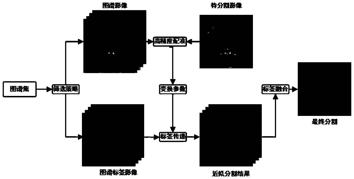
[0076] The present invention will be further described below in conjunction with accompanying drawings and examples. The overall automatic segmentation framework of the liver CT atlas segmentation method based on high-precision registration in the embodiment of the present invention is as follows: figure 2 As shown, the specific implementation s...

Embodiment 2

[0123] The embodiment of the present invention discloses a liver CT atlas segmentation system based on high-precision registration, including:

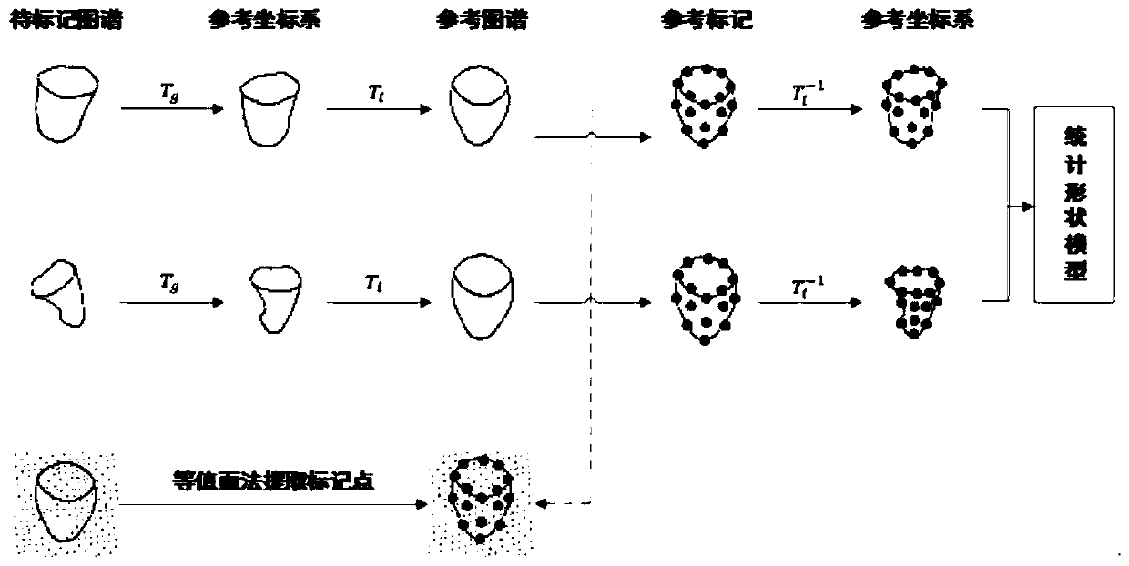
[0124] A model building block for building statistical shape models of all atlases;

[0125] A constraint optimization module, which is used to add the prior information of the statistical shape model to the cost function of the local registration to constrain the mutual information measure;

[0126] The segmentation processing module is used to deform the label image of the atlas to obtain multiple approximate segmentation results by using the inverse array of the deformation field obtained by registration; to perform atlas screening and label fusion on the approximate segmentation results to obtain the final segmentation result.

[0127] The content of Embodiment 1 is also applicable to this system, and will not be repeated here.


PUM


Abstract

The invention discloses a liver CT atlas segmentation method and system based on high-precision registration, which relates to the technical field of medical image processing. The invention firstly performs statistical modeling on the shape of the liver body, thereby restricting the cost function of the local registration and improving the quality of the registration. Then, the labels are mapped by the registered deformation fields to learn the prior shapes of the objects in the maps, and the final segmentation results are obtained. This method can get rid of the inherent limitation of image segmentation, improve the accuracy of segmentation results, and realize fast, accurate and automatic liver segmentation.

Description

technical field [0001] The invention relates to the technical field of medical image processing, in particular to a liver CT atlas segmentation method and system based on high-precision registration. Background technique [0002] Primary liver cancer has a high morbidity and mortality rate in my country. Computed Tomography (CT) technology is widely used in the diagnosis and treatment of liver cancer due to its short scanning time, accurate anatomical information, and high resolution. Accurate extraction of hepatic body tissue is a basic task for computer-aided diagnosis and an important prerequisite for 3D visualization, quantitative analysis, and surgical planning. At present, clinical liver segmentation is still done manually by experience, which is not only time-consuming and laborious, but also has poor repeatability. Therefore, accurate and automatic liver segmentation is of great help to reduce the workload of doctors and improve the speed and accuracy of diagnosis....

Claims


Application Information

Patent Timeline
Patent Type & Authority Patents(China)
IPC IPC(8): G06T7/11G06T7/136G06T7/33
CPCG06T7/11G06T7/136G06T7/344G06T2207/10081G06T2207/30056
Inventor 陆雪松孙鸾
Owner SOUTH CENTRAL UNIVERSITY FOR NATIONALITIES
Why Eureka
Try Eureka