Method and apparatus for attenuation correction

An attenuation correction and attenuation imaging technology, applied in the fields of cardiac imaging and attenuation mapping, can solve the problems of complicated registration process artifacts, and achieve the effect of reducing visible artifacts, accurate and robust attenuation correction

Active Publication Date: 2013-04-03
KONINKLIJKE PHILIPS ELECTRONICS NV
View PDF0 Cites 0 Cited by
  • Summary
  • Abstract
  • Description
  • Claims
  • Application Information

AI Technical Summary

Problems solved by technology

However, these conventional systems are not volumetric (discussed further below), so the imaged subject must be moved during the x-ray imaging procedure in order to image the entire cardiac region
That movement of the object, combined with the object's own movement as it breathes, complicates the registration process and can cause unwanted artifacts in the falloff plot

Method used


Image

  • Method and apparatus for attenuation correction
  • Method and apparatus for attenuation correction
  • Method and apparatus for attenuation correction

Examples


Embodiment Construction

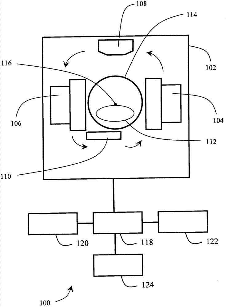
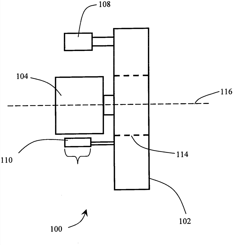
[0017] The imaging methods and apparatus described herein relate generally to any imaging system that generates an attenuation map for another imaging modality, such as PET or SPECT, from data collected by one imaging modality, such as CT. An example of such a device is figure 1 and figure 2 A combined SPECT / CT imaging system 100 is shown. As mentioned above, the imaging methods and apparatuses disclosed herein are applicable to various other kinds of imaging systems.

[0018] Such as figure 1 and figure 2 Illustratively, the system 100 includes a gantry 102 supporting two SPECT gamma-ray detectors 104 and 106 , an X-ray source 108 and a planar X-ray detector 110 . figure 1 A representative object to be imaged, shown at 112 , is partially housed within aperture 114 in gantry 102 . The two gamma-ray detectors 104 and 106 , the X-ray source 108 and the X-ray detector 110 all rotate together and simultaneously on the gantry 102 about an axis of rotation 116 . this is in ...


PUM


Abstract

A method and apparatus of image reconstruction attenuation correction in PET or SPECT cardiac imaging is provided. A volumetric attenuation imaging scan by an X-ray source may be used to generate a gamma ray attenuation map. The volumetric attenuation imaging scan may be randomized, and may be performed while the imaged subject is breathing.

Description

technical field [0001] The present application relates generally to imaging techniques, and more specifically to generating attenuation maps for electronic image reconstruction. This application applies at least to combined PET / CT or SPECT / CT imaging systems, especially for cardiac imaging, and will be described with specific reference thereto. However, the present application also has more general application to generating attenuation maps for other types of imaging, and to other techniques. Background technique [0002] PET (Positron Emission Tomography) and SPECT (Single Photon Emission Computed Tomography) are commonly used to generate diagnostic images of relatively dynamic processes in the human body, such as metabolic activity or blood flow. In PET and SPECT, radioactive isotopes produce gamma rays, which pass through the subject's body and are detected by a gamma-ray detector. Radioisotopes are often administered as radiopharmaceuticals in a subject. Gamma-ray det...

Claims


Application Information

Patent Timeline
Patent Type & Authority Patents(China)
IPC IPC(8): G01T1/161
CPCA61B6/541A61B6/5235A61B6/037A61B6/466A61B6/503G01T1/1611A61B6/032
Inventor J·耶H·梁J·韦塞尔D·索厄德斯-埃梅德L·邵J·L·加勒德
Owner KONINKLIJKE PHILIPS ELECTRONICS NV
Why Eureka
Try Eureka