
Mr-based attenuation correction in pet/mr imaging with dixon pulse sequence

A pulse sequence and imaging technology, applied in the field of medical imaging, can solve the problems of tissue misclassification, long data acquisition time, etc., and achieve the effect of reducing echo time

Active Publication Date: 2016-05-11
KONINKLIJKE PHILIPS NV
View PDF4 Cites 5 Cited by
  • Summary
  • Abstract
  • Description
  • Claims
  • Application Information

AI Technical Summary

Problems solved by technology

[0005] In the Dixon technique, a long TR (repetition time) may be experienced, which results in long data acquisition times
For example, when imaging the location of a whole-body image, the time required to collect all the data exceeds the ability of a typical subject to maintain his or her breathing, leading to image artifacts caused by breathing motion, and thus resulting in Tissue misclassification in the decay map of

Method used


Image

  • Mr-based attenuation correction in pet/mr imaging with dixon pulse sequence
  • Mr-based attenuation correction in pet/mr imaging with dixon pulse sequence

Examples


Embodiment Construction

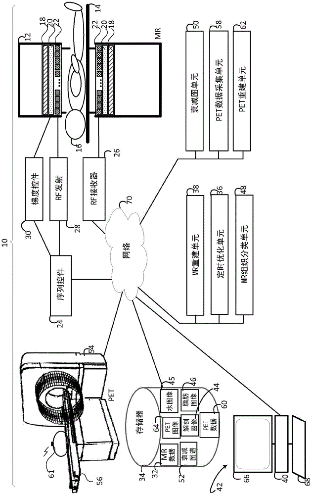
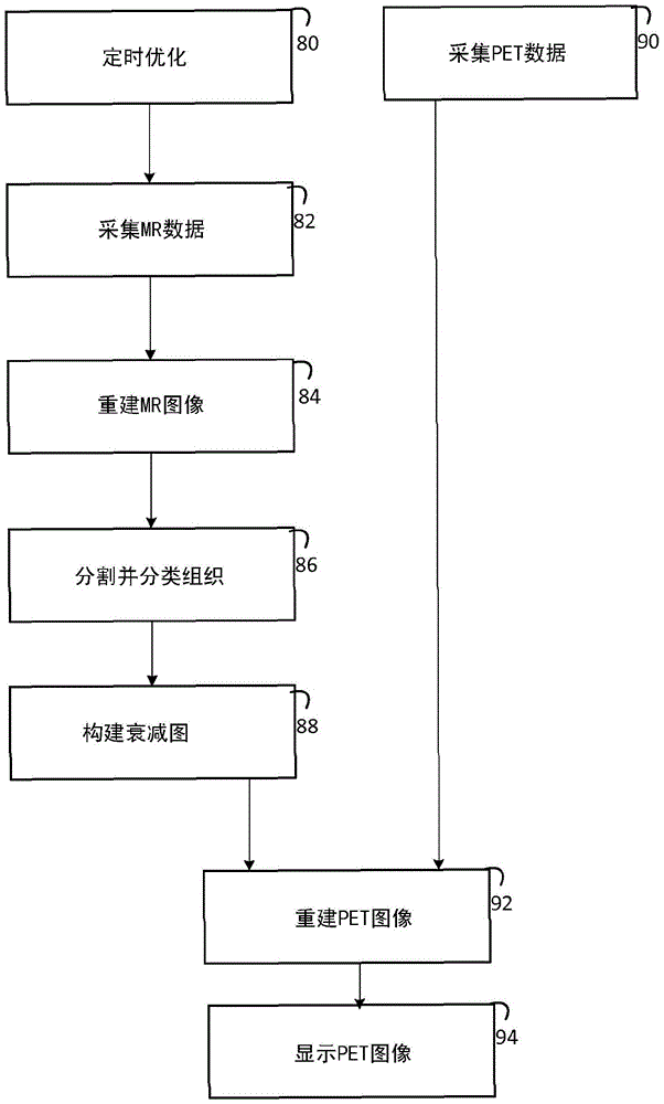
[0027] refer to figure 1 , schematically shows an MR / PET system 10 with timing optimization and MR-based PET attenuation correction maps. System 10 includes a static B shown in partial cross-section 0 MR scanners (or subsystems) 12 of the main magnetic field, such as horizontal bore scanners, open system scanners, c-type scanners, vertical field scanners, hybrid scanners, and the like. The MR scanner includes a subject support 14, such as a horizontal bed or couch, which supports and moves a subject 16 into the MR scanner bore, a static main magnetic field, and a field of view during imaging. The subject support moves the subject to one or more locations that positions a predetermined anatomical portion of the subject within the field of view for imaging. The MR scanner 14 includes a main magnet 18 that generates a static main magnetic field (B 0 ), such as the horizontal main magnetic field. The MR scanner also includes one or more gradient coils 20 for applying gradient ...


PUM


Abstract

A medical imaging system (10) includes a nuclear imaging system (62), a timing optimization unit (40), a magnetic resonance (MR) scanner (12), an MR reconstruction unit (38), and an attenuation map unit (50). The nuclear imaging system (62) receives nuclear decay data and generates at least one nuclear image (64) of a first resolution based on the received nuclear decay data of an imaged subject (16) and an attenuation map (52). The timing optimization unit (40) which selects a first and a second echo time for a modified Dixon (mDixon) pulse sequence and a sufficient number of repetition times (TRs) to generate an image of the subject (16) of at least a first resolution, with the phase angle difference between water and fat at the first and the second echo time being unequal to 0 and 180degrees. The MR scanner (12) applies the sequence to the subject (16) and receives MR data (32) from the subject. The MR reconstruction unit (38) reconstructs at least one MR image (44) based on the MR data (32). The attenuation map unit (50) constructs the attenuation map (52) based on the reconstructed MR image (44).

Description

technical field [0001] The following generally relates to medical imaging. It finds particular application in conjunction with magnetic resonance (MR) imaging to provide attenuation correction for positron emission tomography (PET) imaging, and will be described with particular reference thereto. However, it should be understood that there are also specific applications and other usage scenarios, and are not necessarily limited to the above-mentioned applications. Background technique [0002] In PET imaging, subjects are injected with radiopharmaceuticals that are targeted to specific tissues by uptake, usually based on metabolic activity. When the radiopharmaceutical decays, positrons annihilated in contact with electrons are emitted to form a pair of photons emitted at opposite 180° along the line of response (LOR). The emitted gamma photons are recorded by PET detectors surrounding the object. The location of the annihilation event is calculated from the recorded gamm...

Claims


Application Information

Patent Timeline
Patent Type & Authority Applications(China)
IPC IPC(8): G01R33/48G01R33/561G01R33/56
CPCG01R33/481G01R33/4816G01R33/4828G01R33/5608G01R33/5616G01R33/5615
Inventor C·施特宁H·埃格斯P·博尔纳特L·胡Z·胡
Owner KONINKLIJKE PHILIPS NV
Learn More