
Noise reduction processing method of ultrasound medical image
A technology for medical image and noise reduction processing, which is applied in the field of medical image processing and can solve the problems of medical ultrasound image quality degradation and false structures.
- Summary
- Abstract
- Description
- Claims
- Application Information
AI Technical Summary
Problems solved by technology
Method used
Image

Examples
Embodiment Construction
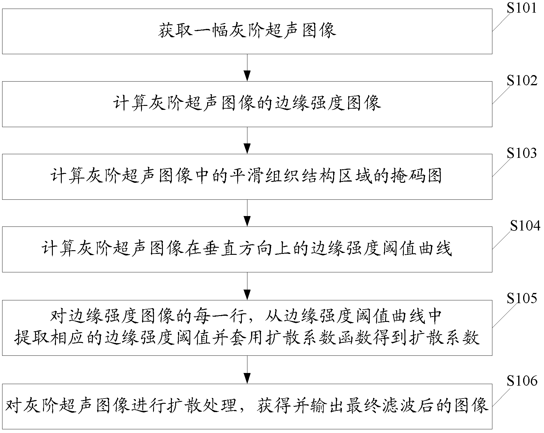
[0072] According to the imaging characteristics of ultrasonic gray-scale images, the present invention uses anisotropic filtering as a framework to propose a dynamic diffusion threshold based on scanning depth to realize a noise reduction method for gray-scale ultrasonic images.
[0073] Such as figure 1 As shown, in a preferred embodiment of the present invention, the noise reduction processing method for gray-scale ultrasound images includes steps:
[0074] Step S101, acquire a gray-scale ultrasound image I of an ultrasound medical image from an image acquisition device or an image storage. If the obtained ultrasonic medical image is not a gray-scale image, it needs to be gray-scaled and processed into a gray-scale ultrasonic image first.
[0075] Step S102, calculating the edge intensity image E of the gray scale ultrasound image I. The edge intensity E of any point coordinate (i, j) on the gray-scale ultrasound image I i,j Represents the possibility that this point is l...
PUM
 Abstract
Description
Claims
Application Information