A Method of Obtaining Initial Contour in Ultrasound Image Segmentation Based on Active Contour Model

An active contour model and ultrasonic image technology, applied in image analysis, image data processing, instruments, etc., can solve the problems of inaccuracy, time-consuming and laborious, etc., and achieve the effect of improving efficiency and accuracy, and clear and accurate initial contour

Inactive Publication Date: 2016-08-17
WUHAN UNIV
View PDF6 Cites 0 Cited by
  • Summary
  • Abstract
  • Description
  • Claims
  • Application Information

AI Technical Summary

Problems solved by technology

[0004] However, the model requires an initial contour. If the initial contour is obtained manually and then converged, it is time-consuming and laborious, and it is not accurate enough.

Method used


Image

  • A Method of Obtaining Initial Contour in Ultrasound Image Segmentation Based on Active Contour Model
  • A Method of Obtaining Initial Contour in Ultrasound Image Segmentation Based on Active Contour Model
  • A Method of Obtaining Initial Contour in Ultrasound Image Segmentation Based on Active Contour Model

Examples


Embodiment 1

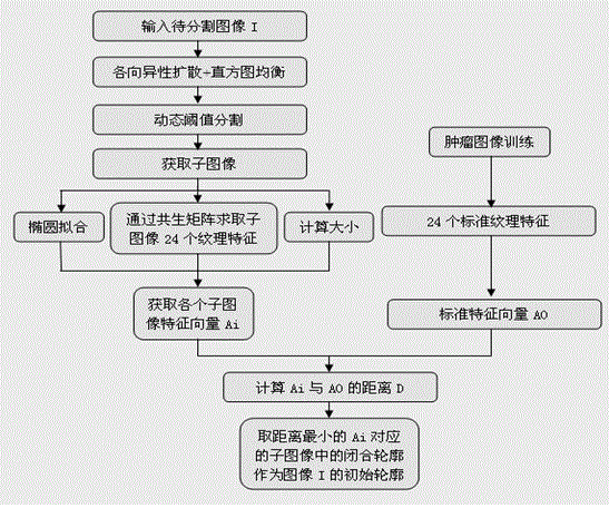
[0029] After training 60 existing liver tumor ultrasound images, the standard feature vector A of liver tumor ultrasound images is obtained 0 . That is, first calculate the 24 texture eigenvalues ​​of each image using the gray level co-occurrence matrix, then 60 groups of eigenvectors containing 24 elements will be obtained, and all eigenvectors will be processed by mathematical linear regression method to obtain a standard The feature vector is to find a vector A with the shortest average distance to the group of feature vectors. The corresponding texture feature values ​​in the vector A are the standard values ​​we need to obtain. Then add the feature vector A to the standard ellipse fitting data 0.7 and the prior size of the liver tumor calculated by the long and short axis of the tumor area marked by the doctor during the ultrasound detection process in the ultrasound image to be tested, and then we get a 26-element standard eigenvector A 0 .

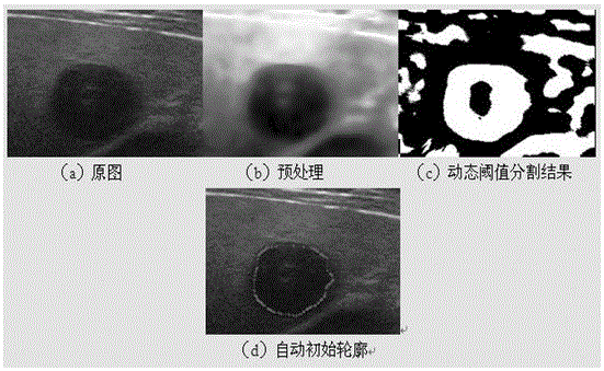
[0030] figure 2 (A) is the ul...

Embodiment 2

[0032] After training 60 ultrasound images of uterine fibroids, the standard feature vector A of uterine fibroids ultrasound images is obtained 0 . That is, first calculate the 24 texture eigenvalues ​​of each image using the gray level co-occurrence matrix, then 60 groups of eigenvectors containing 24 elements will be obtained, and all eigenvectors will be processed by mathematical linear regression method to obtain a standard The feature vector is to find a vector A with the shortest average distance to the group of feature vectors. The corresponding texture feature values ​​in the vector A are the standard values ​​we need to obtain. Then add the feature vector A to the standard ellipse fitting data 0.7 and the prior size of the uterine fibroids calculated from the long and short axis of the tumor area marked by the doctor during the ultrasound detection process in the ultrasound image to be tested, then you get A standard eigenvector A with 26 elements 0 .
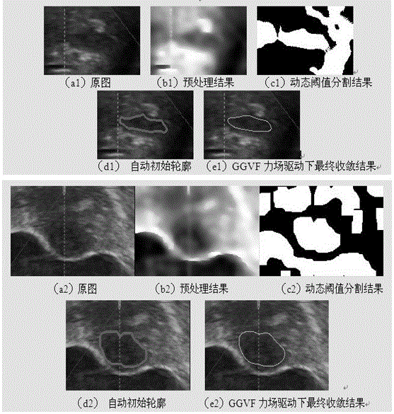
[0033] image 3 ...


PUM


Abstract

The invention discloses a method for acquiring an initial contour in ultrasonic image segmentation based on an active contour model. The method comprises the following steps that the textural features of an ultrasonic image tumor area are trained, and a normal vector is formed by the textural features, a standard elliptical experience value and the tumor prior size in an ultrasonic image to be detected; the ultrasonic image to be detected is preprocessed; dynamic threshold segmentation is carried out on the image; all closed contours generated in the dynamic threshold segmentation result are extracted to form corresponding sub-images; 24 textural features of the sub-images, an ellipse fitting result parameter and the number of pixels in the closed contours of the sub-images are calculated, and a vector is formed by 26 data; the distance between the obtained vector and the normal vector obtained from the tumor area through training is calculated, and the closed contour, corresponding to the vector with the smallest distance, in the sub-images is the contour of a tumor in the segmented ultrasonic image. The method solves the problem that the ultrasonic image has much noise and is fuzzy in boundary, and the initial contour accuracy is high.

Description

Technical field [0001] The invention relates to a method for acquiring an initial contour in an ultrasonic image segmentation based on an active contour, and belongs to the field of ultrasonic image processing. Background technique [0002] With the emergence of high-intensity focused ultrasound, non-invasive treatment of tumors has become more and more widely used. This treatment system has great advantages in clinical practice, such as no surgery, no scars, non-invasive or minimal trauma, and real-time detection and treatment , It is not limited by tumor size, and the total cost is low. These advantages determine that the high-intensity focused ultrasound treatment system has great development and application prospects. The most important thing in the high-intensity focused ultrasound treatment system is to navigate the patient's tumor in real time. The navigation process needs to locate the tumor in the real-time ultrasound image. Changing the traditional manual positioning...

Claims


Application Information

Patent Timeline
Patent Type & Authority Patents(China)
IPC IPC(8): G06T7/00
Inventor 张东龙群芳刘雨周静杨艳
Owner WUHAN UNIV
Why Eureka
Try Eureka