Use of nucleic acid aptamer and its derivatives and rapid imaging method for tissue frozen sections

A technology of frozen section and imaging method, which is applied in the field of pathological diagnosis, can solve the problems of cumbersome steps and long time-consuming, and achieve the effect of simple production steps, less time-consuming and quick judgment

Active Publication Date: 2018-05-29
XIANGYA HOSPITAL CENT SOUTH UNIV
View PDF1 Cites 0 Cited by
  • Summary
  • Abstract
  • Description
  • Claims
  • Application Information

AI Technical Summary

Problems solved by technology

[0006] At present, the detection of specific biomarkers mainly relies on antibodies for immunohistochemical experiments on tissue paraffin sections, which are cumbersome and time-consuming
There are no reports on the use of aptamers and their derivatives for the detection of specific biomarkers in tissue frozen sections for the purpose of tissue imaging

Method used


Image

  • Use of nucleic acid aptamer and its derivatives and rapid imaging method for tissue frozen sections
  • Use of nucleic acid aptamer and its derivatives and rapid imaging method for tissue frozen sections
  • Use of nucleic acid aptamer and its derivatives and rapid imaging method for tissue frozen sections

Examples


Embodiment 1

[0029] Example 1. Imaging Application of EpCAM Aptamer in Frozen Sections of Colon Cancer and Rectal Cancer Tissue

[0030] The rapid imaging method of the nucleic acid aptamer and its derivatives provided by the present invention to tissue frozen sections comprises the following steps:

[0031] 1. Collection of tumor specimens:

[0032] In the operating room, samples are taken from the cancerous tissue mass, the junction of the cancerous tissue mass and normal tissue, and the normal tissue of the same patient, and are marked as: cancerous tissue, junctional tissue, and normal tissue, and then frozen sections are made immediately , used for CY3-labeled SYL3C staining and hematoxylin-eosin (hematoxylin-eosin, HE) section preparation. All specimens were obtained from Xiangya Hospital of Central South University;

[0033] 2. Preparation of tissue frozen sections:

[0034]The specifications of the tissue to be taken are: length × width × thickness = about 24 × 24 × 2mm. Place t...

Embodiment 2

[0055] Example 2. Application of nucleic acid aptamer Adipo8 in specific imaging of frozen section of rat adipose tissue

[0056] The rapid imaging method of the nucleic acid aptamer and derivatives thereof provided by the present invention to tissue frozen sections comprises the following steps:

[0057] 1. Collection of tissue samples:

[0058] All animal specimens were taken from the same rat. Epididymis fat tissue, liver, and skeletal muscle tissue were taken separately, and then frozen sections were made immediately for the production of CY3 fluorescently labeled Adipo-8 staining and HE staining sections; the experimental animals were from the Animal Experiment Center of Xiangya Hospital, Central South University;

[0059] 2, the making of frozen section: as described in embodiment 1;

[0060] 3. Nucleic acid aptamer incubation and HE staining for tissue frozen sections:

[0061] (1) Use a random sequence to block non-specific binding, add 200 microliters of a random s...

experiment example 1

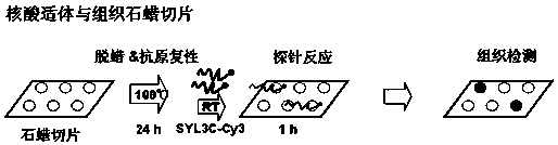
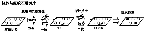
[0069] 1. The imaging results of specific immunostaining with EpCAM nucleic acid aptamer SYL3C on frozen sections of colorectal cancer tissue were compared with the results of specific immunostaining on paraffin sections with SYL3C and commercial EpCAM antibody immunostaining imaging results.

[0070] A series of tissue frozen sections and paraffin sections were made from normal tissues, cancerous tissues, and junctional tissues between normal tissues and cancerous tissues from the same patient with colon cancer and rectal cancer.

[0071] Frozen and paraffin section preparation and HE staining refer to the above method:

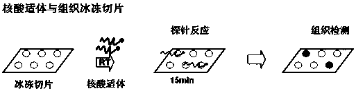
[0072] figure 1 It is the flow chart of EpCAM nucleic acid aptamer immunostaining for frozen tissue sections. The figure shows that the frozen tissue sections are incubated with fluorescent signal CY3-labeled SYL3C for 15 minutes at room temperature and then placed under a fluorescence microscope to observe the imaging results. In order to compare SYL3C wit...


PUM

PropertyMeasurementUnit
particle diameteraaaaaaaaaa

Abstract

The invention discloses application of an aptamer and derivatives of the aptamer and a fast imaging method for frozen tissue slices. The application is to detect specific biomarkers in frozen tissue slices. The method comprises the following steps: blocking the frozen tissue slices by a random sequence and carrying out non-specific binding; then incubating the frozen tissue slices by virtue of a signal-labeled aptamer or an aptamer derivative, observing the imaging results of the frozen tissue slices and judging the location of a focus according to the position of the signal in the frozen tissue slices. The invention provides a new and effective molecular tool for the diagnosis of diseases; and the method is fast, convenient and low in cost.

Description

technical field [0001] The invention belongs to a pathological diagnosis method, and in particular relates to the use of a nucleic acid aptamer and its derivatives and a rapid imaging method for tissue frozen sections. Background technique [0002] After the disease occurs, tissue cells often show a series of pathological changes and express some specific biomarkers. The disease can be diagnosed by identifying the pathological changes of the tissue. The discovery of cell-specific biomarkers is helpful for further diagnosis of diseases, guidance of treatment and judgment of prognosis. Frozen and paraffin sections are most commonly used in clinicopathological work. Compared with paraffin sections, frozen sections are made with simpler steps and are closer to fresh tissues, preserving the structure and even functions of proteins to the greatest extent, without the need for antigen renaturation. But currently, frozen sections are only used for rapid pathological diagnosis unde...

Claims


Application Information

Patent Timeline
Patent Type & Authority Patents(China)
IPC IPC(8): G01N1/28G01N1/30G01N21/64G01N21/65
Inventor 刘慧霞蒲颖张佳妮谭蔚泓陆艺刘真序袁鹏刘俊
Owner XIANGYA HOSPITAL CENT SOUTH UNIV
Why Eureka
Try Eureka