Ultrasound contrast imaging method and region detection and development methods for contrast images

A technology for contrast image and area detection, which is applied in ultrasonic/sonic/infrasound image/data processing, image enhancement, image analysis, etc. It can solve the problem that the phase and amplitude do not meet the linear cancellation conditions, and the contrast signal and tissue residue cannot be distinguished. , linear components cannot be eliminated, etc., to achieve the effect of being beneficial to clinical observation, improving CTR, and reducing inspection costs

Active Publication Date: 2015-06-24
SHENZHEN MINDRAY BIO MEDICAL ELECTRONICS CO LTD +1
View PDF6 Cites 14 Cited by
  • Summary
  • Abstract
  • Description
  • Claims
  • Application Information

AI Technical Summary

Problems solved by technology

[0007] Third, because the signal is too large, each system has its own cancellation performance. After small signal cancellation, the signal is small, and after strong signal cancellation, the tissue residue is also strong. Strong tissue residue makes it impossible to distinguish the angiographic signal from the tissue residue
[0008] Fourth, due to saturation, amplitude modulation is used in the nonlinear detection method, and several pulses with different amplitudes are transmitted. When receiving, the low-amplitude echo signal is readjusted according to the amplitude ratio between the high and low pulses, and compared with the high-amplitude echo signal. Subtraction, which can eliminate the fundamental signal of the tissue; saturation makes the high and low amplitude signals not satisfy the proportional relationship, resulting in the linear component of the tissue that cannot be canceled and remains in the contrast image
[0009] Fifth, due to movement, the phase and amplitude of several transmitted waveforms do not meet the linear cancellation conditions, so that the linear tissue components are eliminated and left in the contrast image
Therefore, in the current contrast-enhanced ultrasound imaging, the existence of tissue residues cannot be avoided

Method used


Image

  • Ultrasound contrast imaging method and region detection and development methods for contrast images
  • Ultrasound contrast imaging method and region detection and development methods for contrast images
  • Ultrasound contrast imaging method and region detection and development methods for contrast images

Examples


Embodiment 1

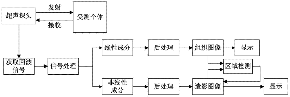
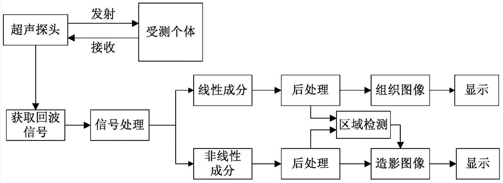
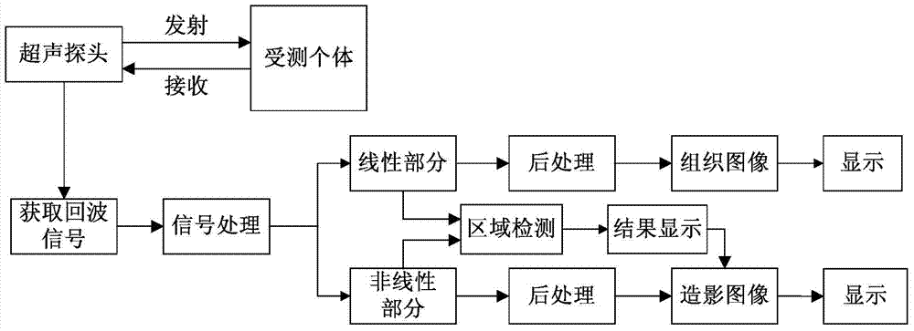
[0044] like Figure 1 to Figure 3 As shown, a contrast-enhanced ultrasound imaging method provided by an embodiment of the present invention specifically includes the following steps:

[0045] S101. Transmit ultrasonic waves to the individual under test through the ultrasonic probe, and obtain echo signals;

[0046] S102. Process the echo signal, and extract linear components and nonlinear components of the echo signal;

[0047] S103. Perform multi-link post-processing on the linear component and nonlinear component of the echo signal, respectively generate tissue images and contrast images, and display them;

[0048] S104. Perform region detection on the contrast image.

[0049] Specifically, in S101, the individual to be tested is generally a human body, and the ultrasonic wave passes through reflection, refraction and scattering (mainly reflection) during the propagation of the human body, and the echo with the anatomical characteristics of human tissue propagates back to...

Embodiment 2

[0056] An embodiment of the present invention provides a region detection method of a contrast image, which is applicable to contrast images formed by nonlinear fundamental wave contrast images, second harmonic contrast images, and any other nonlinear detection techniques.

[0057] like Figure 4 As shown, a region detection method of a contrast image provided by an embodiment of the present invention includes the following steps:

[0058] S201. Acquiring tissue signals and contrast signals during contrast-enhanced ultrasound imaging;

[0059] S202. Preprocessing the tissue signal and the imaging signal respectively;

[0060] S203. Perform comparison processing on the preprocessed tissue signal and the imaging signal, and obtain a histogram distribution diagram of signal values ​​of the image formed after the comparison processing;

[0061] S204. Determine the thresholds of the contrast agent signal and the tissue residual signal through the histogram, and segment the image ...

Embodiment 3

[0073] On the basis of Embodiment 2, the embodiment of the present invention provides a method for developing a contrast image. By controlling the display weights of the contrast agent, tissue residue and noise in the contrast image, the contrast between the contrast agent and tissue residue can be improved, and the The CTR effect of the contrast image can also reduce the display of noise and improve the SNR (Signal to Noise Ratio, signal-to-noise ratio) effect of the contrast image.

[0074] like Image 6 As shown, a method for developing a contrast image provided by an embodiment of the present invention includes using the area detection method for a contrast image described in Embodiment 2 to segment the contrast image into a contrast agent area, a tissue residual area, and a noise area, and also includes : Multiply the contrast agent area, tissue residual area, and noise area by different coefficients P1, P2, and P3 respectively, and then combine them into a new contrast i...


PUM


Abstract

A contrast-enhanced ultrasound imaging method and a method for regional detection and development of a contrasted image. The method for regional detection of a contrasted image comprises: acquiring a tissue signal and a contrast signal in a contrast-enhanced ultrasound imaging process (S201); preprocessing respectively the tissue signal and the contrast signal (S202); comparison processing the preprocessed tissue signal and the contrast signal (S203); by means of a threshold segmentation approach, segmenting an image formed when comparison processed into a contrast agent effect region, a tissue residue effect region, and a noise effect region (S204); and, respectively marking corresponding positions on the contrasted image as a contrast agent region, the tissue residue effect region, and the noise effect region (S205). The regional detection method greatly increases the CTR of a contrast-enhanced ultrasound image, facilitates clinical observation, and enhances the contrast agent signal, thus allowing for a reduced contrast agent dosage injected during contrast-enhanced ultrasound imaging and effectively reduced examination costs.

Description

technical field [0001] The invention relates to ultrasound contrast imaging technology, in particular to an ultrasound contrast imaging method and a region detection and imaging method of contrast images. Background technique [0002] Ultrasound imaging is a relatively mature non-invasive detection method at present. Compared with CT and MRI (Magnetic Resonance Imaging, Magnetic Resonance Imaging, Magnetic Resonance Imaging, magnetic resonance imaging), ultrasonic diagnosis has the advantages of less radiation, low cost, and convenient operation, making ultrasonic diagnosis the primary clinical examination method. [0003] Contrast-enhanced ultrasound is the use of contrast agents to enhance scattered echoes, which can effectively improve the resolution, sensitivity and specificity of ultrasound diagnosis. The core of contrast-enhanced ultrasound imaging technology is how to extract the echo component of the contrast agent in the echo signal and suppress the echo component o...

Claims


Application Information

Patent Timeline
Patent Type & Authority Applications(China)
IPC IPC(8): A61B8/08A61B8/00
CPCG01S7/52077A61B8/481A61B8/5207G01S7/52039G06T7/11G06T7/136G06T2207/10132G06T2207/30004A61B8/08A61B8/5269A61B8/5292
Inventor 储霞桑茂栋
Owner SHENZHEN MINDRAY BIO MEDICAL ELECTRONICS CO LTD
Why Eureka
Try Eureka