Method for contrast-enhanced ultrasound imaging and region detection and imaging method for contrast-enhanced images
A technology for contrast image and area detection, which is applied in ultrasonic/acoustic/infrasonic image/data processing, image enhancement, image analysis, etc. It can solve the problem that the phase and amplitude do not meet the linear cancellation condition, and the contrast signal and tissue residue cannot be distinguished , linear components cannot be eliminated, etc., to achieve the effect of facilitating clinical observation, improving CTR, and reducing inspection costs
- Summary
- Abstract
- Description
- Claims
- Application Information
AI Technical Summary
Problems solved by technology
Method used
Image
Examples
Embodiment 1
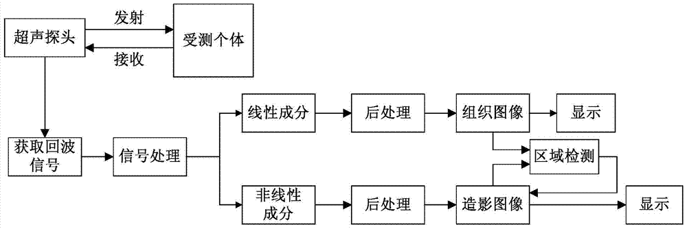
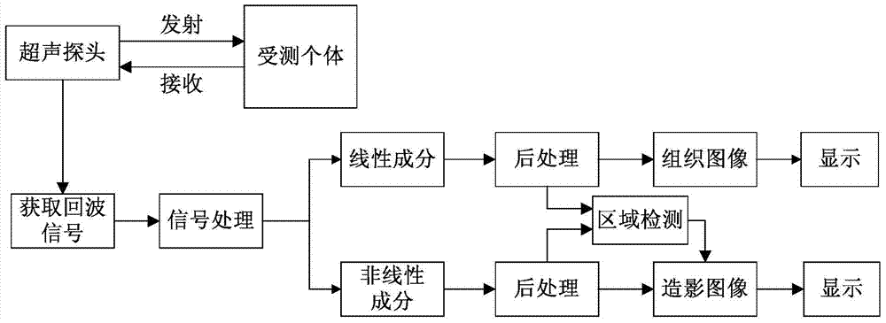
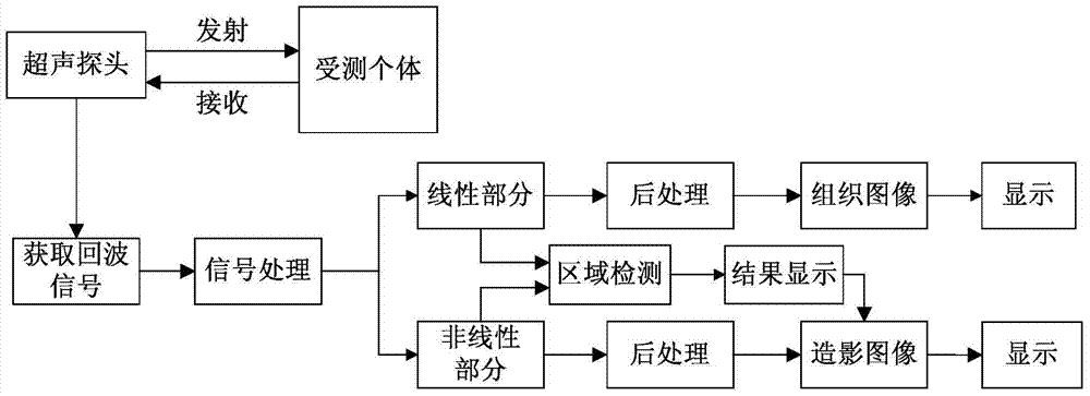
[0044] Such as Figure 1 to Figure 3 As shown, a contrast-enhanced ultrasound imaging method provided by an embodiment of the present invention specifically includes the following steps:
[0045] S101. Transmit ultrasonic waves to the individual under test through the ultrasonic probe, and obtain echo signals;
[0046] S102. Process the echo signal, and extract linear components and nonlinear components of the echo signal;
[0047] S103. Perform multi-link post-processing on the linear component and nonlinear component of the echo signal, respectively generate tissue images and contrast images, and display them;
[0048] S104. Perform region detection on the contrast image.
[0049] Specifically, in S101, the individual to be tested is generally a human body, and the ultrasonic wave passes through reflection, refraction and scattering (mainly reflection) during the propagation of the human body, and the echo with the anatomical characteristics of human tissue propagates back...
Embodiment 2
[0056] An embodiment of the present invention provides a region detection method of a contrast image, which is applicable to contrast images formed by nonlinear fundamental wave contrast images, second harmonic contrast images, and any other nonlinear detection techniques.
[0057] Such as Figure 4 As shown, a region detection method of a contrast image provided by an embodiment of the present invention includes the following steps:
[0058] S201. Acquiring tissue signals and contrast signals during contrast-enhanced ultrasound imaging;
[0059] S202. Preprocessing the tissue signal and the imaging signal respectively;
[0060] S203. Perform comparison processing on the preprocessed tissue signal and the imaging signal, and obtain a histogram distribution diagram of signal values of the image formed after the comparison processing;
[0061] S204. Determine the thresholds of the contrast agent signal and the tissue residual signal through the histogram, and segment the ima...
Embodiment 3
[0073] On the basis of Embodiment 2, the embodiment of the present invention provides a method for developing a contrast image. By controlling the display weights of the contrast agent, tissue residue and noise in the contrast image, the contrast between the contrast agent and tissue residue can be improved, and the The CTR effect of the contrast image can also reduce the display of noise and improve the SNR (Signal to Noise Ratio, signal-to-noise ratio) effect of the contrast image.
[0074] Such as Figure 6 As shown, a method for developing a contrast image provided by an embodiment of the present invention includes using the area detection method for a contrast image described in Embodiment 2 to segment the contrast image into a contrast agent area, a tissue residual area, and a noise area, and also includes : Multiply the contrast agent area, tissue residual area, and noise area by different coefficients P1, P2, and P3 respectively, and then combine them into a new contra...
PUM
 Abstract
Description
Claims
Application Information
 - R&D
- Intellectual Property
- Life Sciences
- Materials
- Tech Scout
- Unparalleled Data Quality
- Higher Quality Content
- 60% Fewer Hallucinations
Browse by: Latest US Patents, China's latest patents, Technical Efficacy Thesaurus, Application Domain, Technology Topic, Popular Technical Reports.
© 2025 PatSnap. All rights reserved.Legal|Privacy policy|Modern Slavery Act Transparency Statement|Sitemap|About US| Contact US: help@patsnap.com


