Truncated Dirichlet process infinite Student'st' hybrid model-based brain nuclear magnetic resonance image segmentation method

A technology of nuclear magnetic resonance image and hybrid model, which is applied in image analysis, image enhancement, image data processing and other directions, and can solve the problems of noise sensitivity, inability to automatically determine the number of divisions, and complex sampling calculation.

CN106023236AActive Publication Date: 2016-10-12HUAQIAO UNIVERSITY
2 Cites 5 Cited by

Patent Information

Authority / Receiving Office
CN · China
Patent Type
Applications(China)
Current Assignee / Owner
Publication Date
2016-10-12

Smart Images

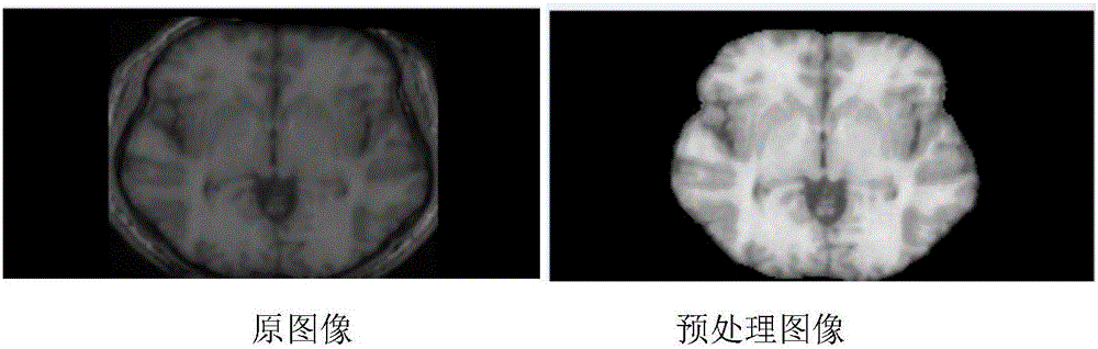
  • Figure 1
    Figure 1
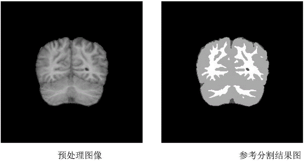
  • Figure 2
    Figure 2
  • Figure 3
    Figure 3
Patent Text Reader

Abstract

The invention relates to a truncated Dirichlet process infinite Student'st' hybrid model-based brain nuclear magnetic resonance image segmentation method. The method includes the following steps that: based on a Dirichlet process infinite Student'st' hybrid model, a component number in the infinite Student'st' hybrid model is assumed to be a preset image segmentation number K; an expectation maximization algorithm is adopted to solve the model; and image segmentation is carried out by using a Bayesian maximum posterior probability criterion. According to the method of the invention, the assumed Student'st' hybrid model directly corresponds to different portions of a brain nuclear magnetic resonance image; the high tail characteristics of Student'st' distribution determines that the model has a good anti-noise effect, so that the segmentation of the brain nuclear magnetic resonance image can be realized; in the processing of solving the Dirichlet process infinite Student'st' hybrid model, the simple and efficient expectation maximization algorithm is adopted to solve the Dirichlet process infinite Student'st' hybrid model, and the solving of the model is easier to realized; and one brain nuclear magnetic resonance image can be quickly and automatically segmented at a PC end.
Need to check novelty before this filing date? Find Prior Art

Description

technical field

[0001] The present invention relates to computer-aided image processing technology, more specifically, relates to a kind of brain MRI image segmentation method based on truncated Dirichlet process infinite Student's t mixture model. Background technique

[0002] In recent decades, medical imaging technology has developed rapidly. In particular, magnetic resonance imaging technology has the advantages of non-invasiveness, inspection range covering various systems of the human body, and rich imaging data. It is the most widely used. Medical image analysis methods have been concerned, researched and applied. Medical image segmentation is an important research content in medical image analysis.

[0003] Medical image segmentation is to divide the image into several regions according to the similarity in the medical image region and the different characteristics between regions, mainly including studying anatomical structures, identifying regions of interest, obse...

Examples

Embodiment Construction

[0055] The present invention will be further described in detail below in conjunction with the accompanying drawings and embodiments.

[0056] In order to solve the deficiencies in the prior art, the present invention provides a brain MRI image segmentation method based on the truncated Dirichlet process infinite Student's t mixture model, so that the user can obtain the segmentation result of the brain MRI image simply and quickly.

[0057] The present invention is based on the infinite Student's t mixture model of the Dirichlet process, considering that the infinite Student's t mixture model of the Dirichlet process is actually countably infinite, therefore, the component number in the infinite Student's t mixture model is assumed to be the division number K of the preset image (an appropriate rather large number K), and then use the expectation maximization algorithm to solve the model, and then use the Bayesian maximum posterior probability criterion to perform image segmen...