Identification method of primary central nervous system lymphoma and glioblastoma based on sparse representation system

A glioblastoma, sparse representation technology, used in character and pattern recognition, computer parts, image data processing, etc.

Active Publication Date: 2017-08-04
FUDAN UNIV
View PDF7 Cites 22 Cited by
  • Summary
  • Abstract
  • Description
  • Claims
  • Application Information

AI Technical Summary

Problems solved by technology

Some advanced MRI imaging methods such as diffusion-weighted imaging and dynamic susceptibility-enhanced perfusion imaging have been used to assis

Method used


Image

  • Identification method of primary central nervous system lymphoma and glioblastoma based on sparse representation system
  • Identification method of primary central nervous system lymphoma and glioblastoma based on sparse representation system
  • Identification method of primary central nervous system lymphoma and glioblastoma based on sparse representation system

Examples


Embodiment Construction

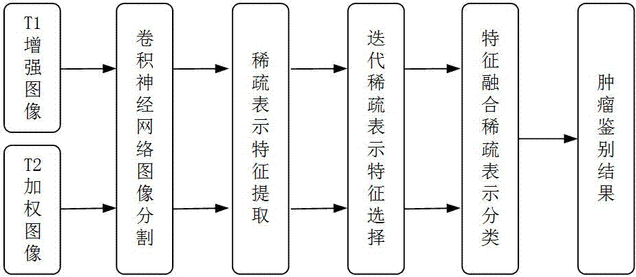
[0044] The following are the specific implementation steps of the whole method:

[0045] 1. Firstly, perform brain removal and gray-scale normalization operations on the images in the data set, and select 40 images from the T1-enhanced images and T2-weighted image sets for manual labeling of tumor areas, and then send the labeling results and corresponding images to Two kinds of convolutional neural networks constructed were used to train the network parameters, and finally the two trained convolutional neural networks were used to segment the tumor area of ​​the corresponding modality image.

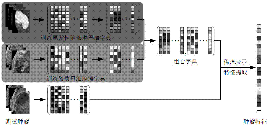
[0046] 2. Extract the set of image blocks contained in the tumor area, the size of the image blocks is 11*11, and the center interval of the image blocks is 5*5. For T1-enhanced modal images, select the image block sets corresponding to 20 cases of primary brain lymphoma images, use the K singular value decomposition method to train the primary brain lymphoma dictionary, and select 20 c...


PUM


Abstract

The invention belongs to the technical field of computer auxiliary diagnosis, and specifically relates to an identification method of primary central nervous system lymphoma and glioblastoma based on a sparse representation system. The method includes: segmenting T1 enhanced and T2 weighted MRI image tumor regions by employing an image segmentation method based on a convolutional neural network; then designing a dictionary learning and sparse representation method, and extracting texture characteristics of the tumor regions; selecting some characteristics with high stability and high resolution for tumor identification by employing an iterative sparse representation characteristic selection method in order to reduce the characteristic redundancy and improve the tumor identification efficiency; and finally establishing a combined sparse representation classification model containing two modals of T1 enhanced or T2 weighted based on the thought of eigenstate fusion in order to improve the tumor identification precision. According to the method, high tumor identification precision can be obtained, manual operation for extraction of identification parameters is avoided, the robustness is high, and the method can be applied to clinic identification of primary central nervous system lymphoma and glioblastoma.

Description

technical field [0001] The invention belongs to the technical field of computer-aided diagnosis, in particular to a method for distinguishing primary brain lymphoma and glioblastoma based on a sparse representation system. Background technique [0002] Clinically, there are large differences in the treatment options for primary central nervous system lymphoma (PCNSL) and glioblastoma (GBM). Accurate identification of the two before treatment can guide clinicians Making a reasonable treatment plan has important clinical value. However, it is very difficult to accurately distinguish primary brain lymphoma from glioblastoma using some traditional modal MRI images such as T1-weighted, T1-enhanced, T2-weighted, and T2FLAIR, because the images of these two tumors in these modalities exhibit many similar properties. Some advanced MRI imaging methods such as diffusion-weighted imaging and dynamic susceptibility-enhanced perfusion imaging have been used to assist in the diagnosis o...

Claims


Application Information

Patent Timeline
IPC IPC(8): G06K9/46G06K9/62G06T7/00
CPCG06T7/0012G06T2207/10088G06T2207/30096G06V10/424G06V10/513G06F18/253
Inventor 汪源源余锦华吴国庆李泽榉
Owner FUDAN UNIV
Why Eureka
Try Eureka