Tumor cell phenotype recognition counting method based on cell fluorescence image

A fluorescence image, tumor cell technology, applied in the field of image analysis, can solve the problems of inability to count, unable to achieve fully automatic effect, manual adjustment of system parameters, etc., to achieve the effect of small error, shortened recognition counting time, and high recognition accuracy

A fluorescence image, tumor cell technology, applied in the field of image analysis, can solve the problems of inability to count, unable to achieve fully automatic effect, manual adjustment of system parameters, etc., to achieve the effect of small error, shortened recognition counting time, and high recognition accuracy

CN111862004AActive Publication Date: 2020-10-30JIANGXI UNIVERSITY OF TRADITIONAL CHINESE MEDICINE

Image

  • Tumor cell phenotype recognition counting method based on cell fluorescence image
  • Tumor cell phenotype recognition counting method based on cell fluorescence image
  • Tumor cell phenotype recognition counting method based on cell fluorescence image

Examples


Embodiment Construction

[0036] The present invention will be further described below in conjunction with the embodiments and accompanying drawings.

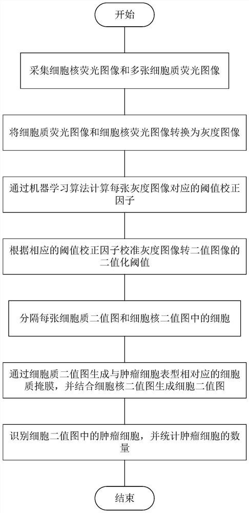
[0037] Such as figure 1 The flow chart of the tumor cell phenotype recognition and counting method based on the cell fluorescence image shown, the recognition calculation method includes:

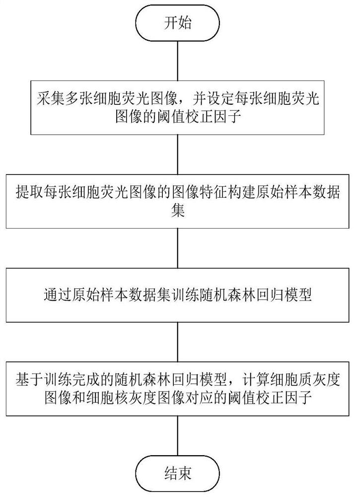
[0038] Step 1. Acquire multiple nuclear fluorescence images and cytoplasmic fluorescence images. The fluorescence image of the cell nucleus is a fluorescence image collected after the cells are stained with a cell nucleus marker, such as the nucleus staining reagent DAPI. Cytoplasmic fluorescence images are fluorescent images collected after cells are stained with different types of cytoplasmic markers, such as leukocyte markers-leukocyte common antigen, tumor-specific protein markers-keratin or cell surface vimentin. The collected cytoplasmic fluorescence images correspond one-to-one with the cytoplasmic markers used. In this way, a variety of tumor cells with di...


PUM


Abstract

The invention discloses a tumor cell phenotype recognition counting method based on a cell fluorescence image, and the method comprises the steps: collecting a cell nucleus fluorescence image and a plurality of cytoplasm fluorescence images, and converting the fluorescence images into a gray level image; determining a threshold correction factor of each grayscale image through a machine learning algorithm; correcting a binarization threshold value in a process of converting the grayscale image into the binary image according to a threshold value correction factor; generating a cytoplasm mask corresponding to a tumor cell phenotype through a cytoplasm binary image, generating a cell binary image in combination with a cell nucleus binary image, finally, recognizing tumor cells in the cell binary image, and counting the number of the tumor cells. Two methods based on rules and statistical machine learning are combined, various circulating tumor cell phenotypes can be fully automatically and flexibly recognized and counted under different image conditions, the recognition precision is high, counting is accurate, errors are small, and the recognition counting time is short.

Description

technical field [0001] The invention relates to the field of image analysis, in particular to a method for identifying and counting tumor cell phenotypes based on cell fluorescence images. Background technique [0002] Circulating tumor cells (CTCs) are tumor cells that shed from the original tumor and enter the blood circulation, and their counting has clinical oncological significance such as diagnosis, prognosis, and drug resistance prediction. Circulating tumor cells are often identified by immunofluorescence, typically using staining for nuclei, leukocytes, and tumor-specific protein markers, and are defined as nuclei positive for tumor-specific protein markers but negative for leukocyte markers. [0003] In addition, the heterogeneity of circulating tumor cells contributes to these cells having different phenotypes and expressing different tumor-specific proteins, for example, epithelial circulating tumor cells express cytokeratin and epithelial cell adhesion molecule ...

Claims


Application Information

Patent Timeline
30 Oct 2020
Publication
CN111862004A
IPC
G06T7/00; G06T7/90; G06T7/136; G06T5/00; G06K9/62
CPC
G06T7/0012; G06T7/90; G06T7/136; G06T2207/30024; G06T2207/30242; G06F18/24323; G06T5/70
Inventors
郑国旋; 韩平畴