Femoral head localization method, system, device and storage medium in ultrasound image

A positioning method and technology of the femoral head, applied in the field of images, can solve the problems such as the inability to visually display the femoral head and the bottom and top structures of the acetabulum, the poor display of cartilage and soft tissue structures, the long scanning time and the appointment period, etc., so as to achieve easy programming. Achieving, measuring, and avoiding human-biased results

Active Publication Date: 2022-07-01
BEIJING JISHUITAN HOSPITAL
View PDF5 Cites 0 Cited by
  • Summary
  • Abstract
  • Description
  • Claims
  • Application Information

AI Technical Summary

Problems solved by technology

The above-mentioned technologies all have defects: for example, X-ray and CT can not show cartilage and soft tissue structures well
MRI scanning time and appointment period are longer, and the price is more expensive
Ultrasound of the front of the hip is a cross-sectional image, which cannot directly display the structure of the femoral head and the bottom and top of the acetabulum, and judge the position of the femoral head relative to the acetabulum, so parameter measurement cannot be applied

Method used


Image

  • Femoral head localization method, system, device and storage medium in ultrasound image
  • Femoral head localization method, system, device and storage medium in ultrasound image
  • Femoral head localization method, system, device and storage medium in ultrasound image

Examples


Embodiment approach

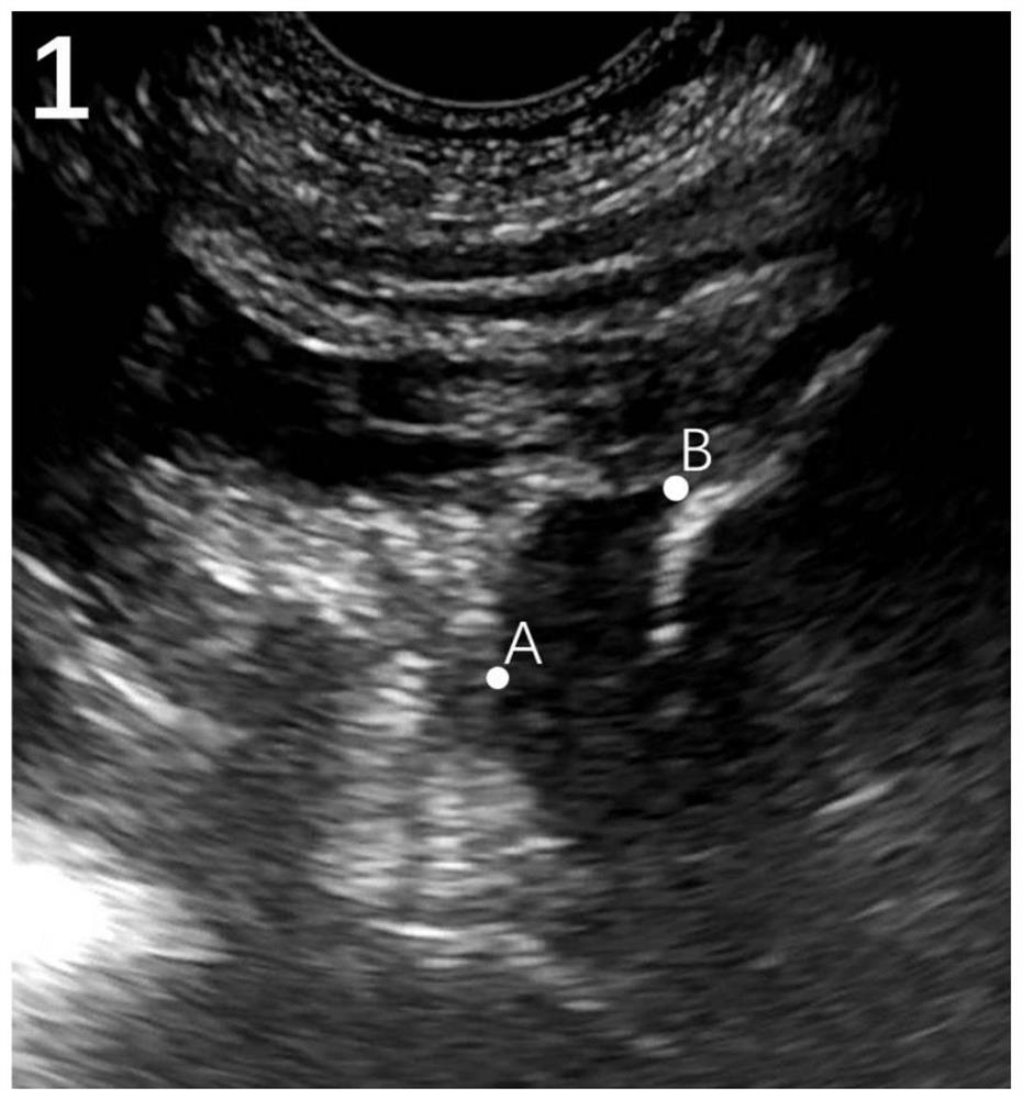
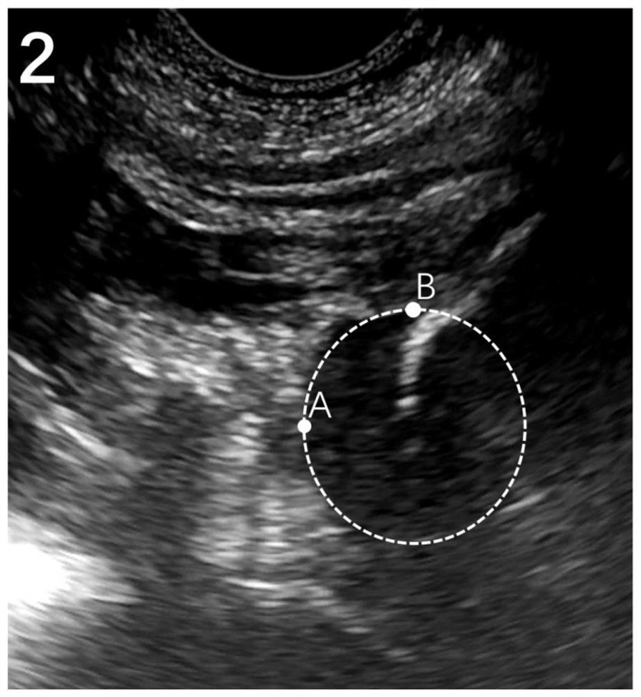
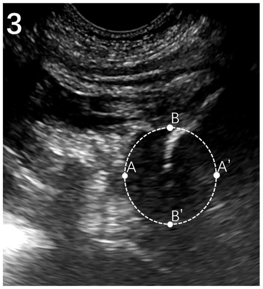
[0070] Embodiments of the present invention provide a method, system, device and storage medium for locating a femoral head in an ultrasound image. This method uses the image of the median coronal section of the acetabulum to calculate the distance L2 between the femoral head and the bottom of the acetabulum, the distance L3 between the femoral head and the center of the acetabulum, and the distance L4 between the femoral head and the top of the acetabulum, which can efficiently and conveniently determine the position of the femoral head. to locate.

[0071] like Figure 23 As shown in the figure, the femoral head positioning method includes: selecting the acetabular median coronal section as the target section, selecting five base points A, B, C, D and E in the acetabular median coronal section image, and using the five base points to measure the femoral head respectively The bone diameter L1, the distance between the femoral head and the bottom of the acetabulum L2, the dis...


PUM


Abstract

The invention discloses a femoral head positioning method, system, device and storage medium in an ultrasonic image. The method includes: respectively measuring the femoral head diameter L1 and the distance L2 between the femoral head and the bottom of the acetabulum in an acetabulum median coronal section image. , The distance between the femoral head and the center of the acetabulum is L3 and the distance between the femoral head and the top of the acetabulum is L4. According to the distance between the femoral head and the bottom of the acetabulum L2, the distance between the femoral head and the center of the acetabulum L3, and the distance between the femoral head and the top of the acetabulum L4, the relative femoral head is determined. The position of the acetabulum can efficiently and conveniently locate the position of the femoral head.

Description

technical field [0001] The invention belongs to the field of image technology, and in particular relates to a method, system, device and storage medium for locating a femoral head in an ultrasonic image. Background technique [0002] The position of the femoral head is mostly observed based on X-ray, CT, MRI and ultrasound images of the anterior hip. All of the above techniques have shortcomings: for example, X-ray and CT do not show cartilage and soft tissue structures well. MRI scan time and appointment period are longer and more expensive. Ultrasound of the front of the hip is a cross-sectional imaging, which cannot visually display the femoral head and the bottom and top structures of the acetabulum, and judge the position of the femoral head relative to the acetabulum, so parametric measurements cannot be used. SUMMARY OF THE INVENTION [0003] The purpose of the present invention is to provide a femoral head positioning method, system, device and storage medium in ...

Claims


Application Information

Patent Timeline
Patent Type & Authority Patents(China)
IPC IPC(8): A61B8/08
CPCA61B8/0875A61B8/5215
Inventor 于静淼
Owner BEIJING JISHUITAN HOSPITAL
Why Eureka
Try Eureka