Method of improving the quality of a three-dimensional ultrasound doppler image

a three-dimensional ultrasound and image technology, applied in image enhancement, instruments, medical/anatomical pattern recognition, etc., can solve the problems of affecting the quality of the image, requiring a long diagnostic time, and limited use of the image, so as to improve the quality of the three-dimensional (3d) ultrasound doppler image

Inactive Publication Date: 2006-08-17
KOREA ADVANCED INST OF SCI & TECH
View PDF10 Cites 20 Cited by
  • Summary
  • Abstract
  • Description
  • Claims
  • Application Information

AI Technical Summary

Benefits of technology

[0004] It is an object of the present invention to provide a method of improving the quality of a three-dimensional (3D) ultrasound Doppler image by applying a diffusion matrix and gradients of voxels to an original ultrasound image.

Problems solved by technology

However, since the method of increasing the transmission power level may produce a certain effect to organs or tissues of the target object, its use is limited.
Also, there are problems in that the method of injecting the contrast medium may harm a blood vessel and require a long diagnostic time due to a contrast agent injection time.

Method used


Image

  • Method of improving the quality of a three-dimensional ultrasound doppler image
  • Method of improving the quality of a three-dimensional ultrasound doppler image
  • Method of improving the quality of a three-dimensional ultrasound doppler image

Examples


Embodiment Construction

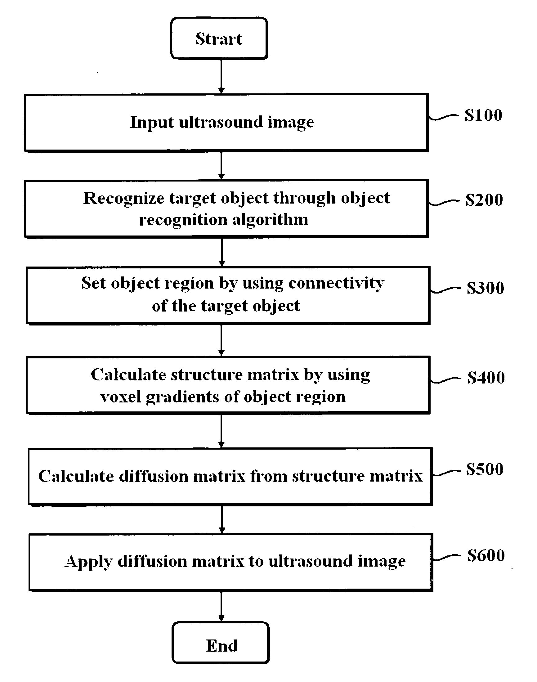
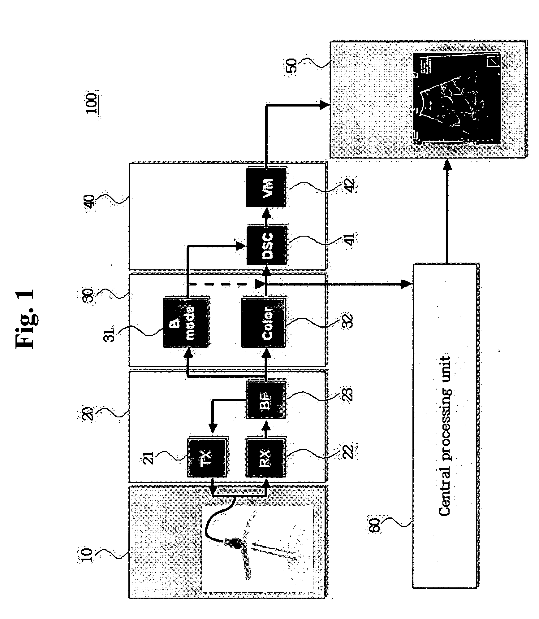
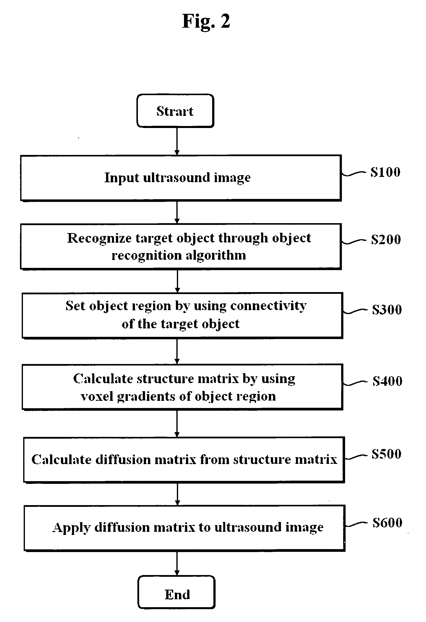
[0018]FIG. 1 is a schematic diagram showing an ultrasound diagnostic system. An ultrasound diagnostic system 100 includes an ultrasound detecting unit 10, a front-end unit 20, an image processing unit 30, a back-end unit 40 and a central processing unit 60.

[0019] The ultrasound detecting unit 10 contains an ultrasound probe. The ultrasound probe has an ultrasound transducer array consisting of a plurality of transducers.

[0020] The front-end unit 20 includes a transmitting unit 21, a receiving unit 22 and a beam forming unit 23. The transmitting unit 21 provides a transmission signal formed in beam forming unit 23 to the probe of the ultrasound detecting unit 10. The receiving unit 22 transmits an ultrasound echo signal received from the probe to the beam forming unit 23.

[0021] The image processing unit 30 includes a B-mode processing unit 31 and a color processing unit 32 for performing image processing upon a reception beam outputted from the beam forming unit 23.

[0022] The bac...


PUM


Abstract

The present invention relates to a method of improving a 3D ultrasound color Doppler image through a post-processing. A method of processing an ultrasound image, includes the following steps: a) recognizing a target object from an inputted ultrasound image based on an object recognition algorithm using connectivity of the target object; b) setting at least one object region by using the connectivity of the recognized target object; c) calculating a structure matrix by using voxel gradients of the object region; d) calculating a diffusion matrix from the structure matrix; and e) acquiring a processed ultrasound image by applying the diffusion matrix and the voxel gradients to the inputted ultrasound image.

Description

FIELD OF THE INVENTION [0001] The present invention generally relates to a method of processing an ultrasound image, and more particularly to a method of improving the quality of a three-dimensional (3D) ultrasound Doppler image by applying post-processing to an ultrasound image acquired through an ultrasound device. BACKGROUND OF THE INVENTION [0002] The ultrasound diagnostic device has several advantages such as being convenient and ensuring safety from exposure to X-rays, etc. For this reason, the ultrasound diagnostic device is extensively utilized in various medical fields. The ultrasound diagnostic device acquires ultrasound images of organs from a target object by using the ultrasound characteristics such as reflection, scattering and absorption, which affect the ultrasound signal emitted into the organs of the target object. The scattered ultrasound signal includes information related to an acoustic impedance difference at boundaries of the organs. Furthermore, the scattered...

Claims


Application Information

Patent Timeline
Patent Type & Authority Applications(United States)
IPC IPC(8): A61B8/00G06T1/00G06T15/08G06V10/30
CPCG06K9/40G06K2209/05G06T5/002G06T5/20G06T2207/10024G06T2207/10136G06T2207/30101G06V10/30G06V2201/03A47G23/04B65D81/18
Inventor KIM, CHEOL ANRA, JONG BEOMSONG, YOUNG SEUKLIM, JUNG EUNYANG, EUN HOYU, DONGHOONLEE, JAE KEUN
Owner KOREA ADVANCED INST OF SCI & TECH
Why Eureka
Try Eureka