Converting large amounts
raw data including
raw data on digital images generated in these experiments into meaningful information that can be used by an analyst to formulate an opinion remains a challenge that hinders many investigators.
Since, cancer is life threatening when it becomes invasive, it carries a potential for spreading and
metastasis.
Clinical studies in patients with
breast cancer over the last decade have convincingly demonstrated that amplification /
over expression of HER-2 / neu is associated with a poor medical prognosis.
Gene amplification of HER-2 / neu is associated with aggressive cell behavior and
poor prognosis.
Prostate cancer strikes as many men (and causes almost as many deaths annually) as
breast cancer does in women, but lacks the national awareness and research funding
breast cancer currently receives.
However, many images do not have pixel values that make effective use of the full
dynamic range of pixel values available on an
output device.
The result in either case is that the output is relatively dull in appearance.
There are several problems associated with using existing
digital image analysis techniques for analyzing digital images for determining know medical conditions.
One problem is that existing
digital image analysis techniques are typically used only for analyzing measurements of chemical compounds applied to biological samples such as groups of cells from a
tissue sample.
Another problem is the manual method used by pathologists is
time consuming and prone to error including missing areas of the slide including tumor or cancer cells.
There is a considerable gap between the pace at which
drug discovery is progressing and automated tools available to assist the
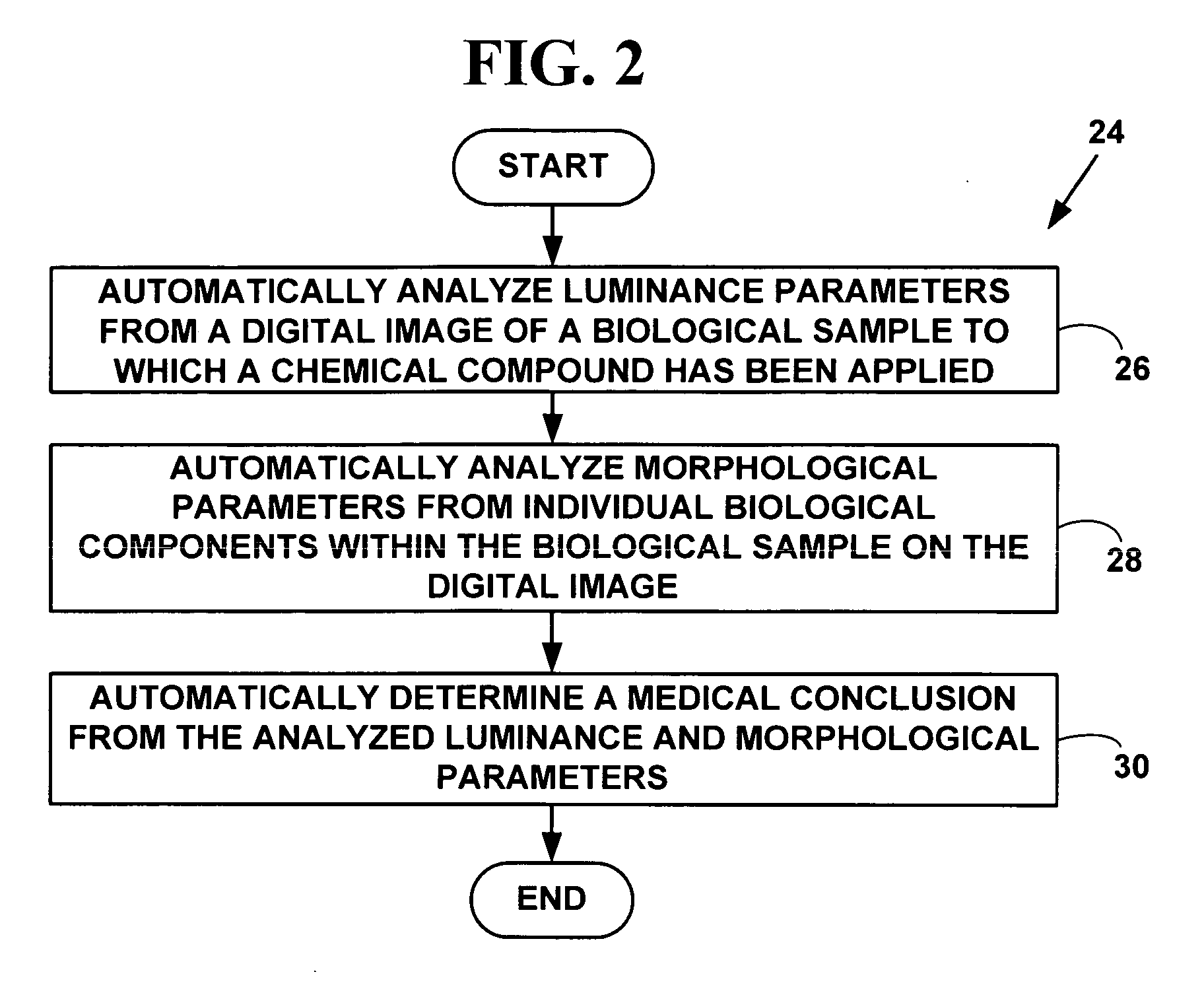
drug researcher.
Often researchers are forced to resort to manual methods which are subjective,
time consuming and could be inconsistent.
Difficulties are reported with the performance and interpretation of IHC and FISH in the medical
oncology community over the last several years.
In particular the detection of
estrogen and progesterone receptors and the detection of HER2 / neu targets is not consistent.
Problems encountered include, but are not limited to, identification of cutoff points in
assay scores to define positive vs. negative results, broad interlaboratory variability in performance of the assays, discrepancies between laboratories with high volume vs
low volume throughput, use of “home brew” antibodies for IHC, deviations from recommended methods in the
package insert leading to altered performance characteristics of the assays, conflicting data in the published literature, and lack of data from prospectively conducted studies.
There are several problems associated with using existing
digital image analysis techniques for analyzing FISH images.
Once there are a large number of image sections to be processed together, issues like seamless composition of tiles becomes an issue.
3) Another problem with FISH based analysis is the need to stack planes in three dimensions to get a focused image.
Failure to obtain interpretable results after two test runs occurred in 5.0% of samples tested by FISH and 0.08% of those tested by
immunohistochemistry.
On the website(www.ihcworld.com / _protocols / general_IHC / immunoenzyme_double.htm), it is stated that this
enzyme based
double staining method is limited for the demonstration of two proteins at different locations.
Often, the tissue origin of a tumor cannot be determined with routine
histology.
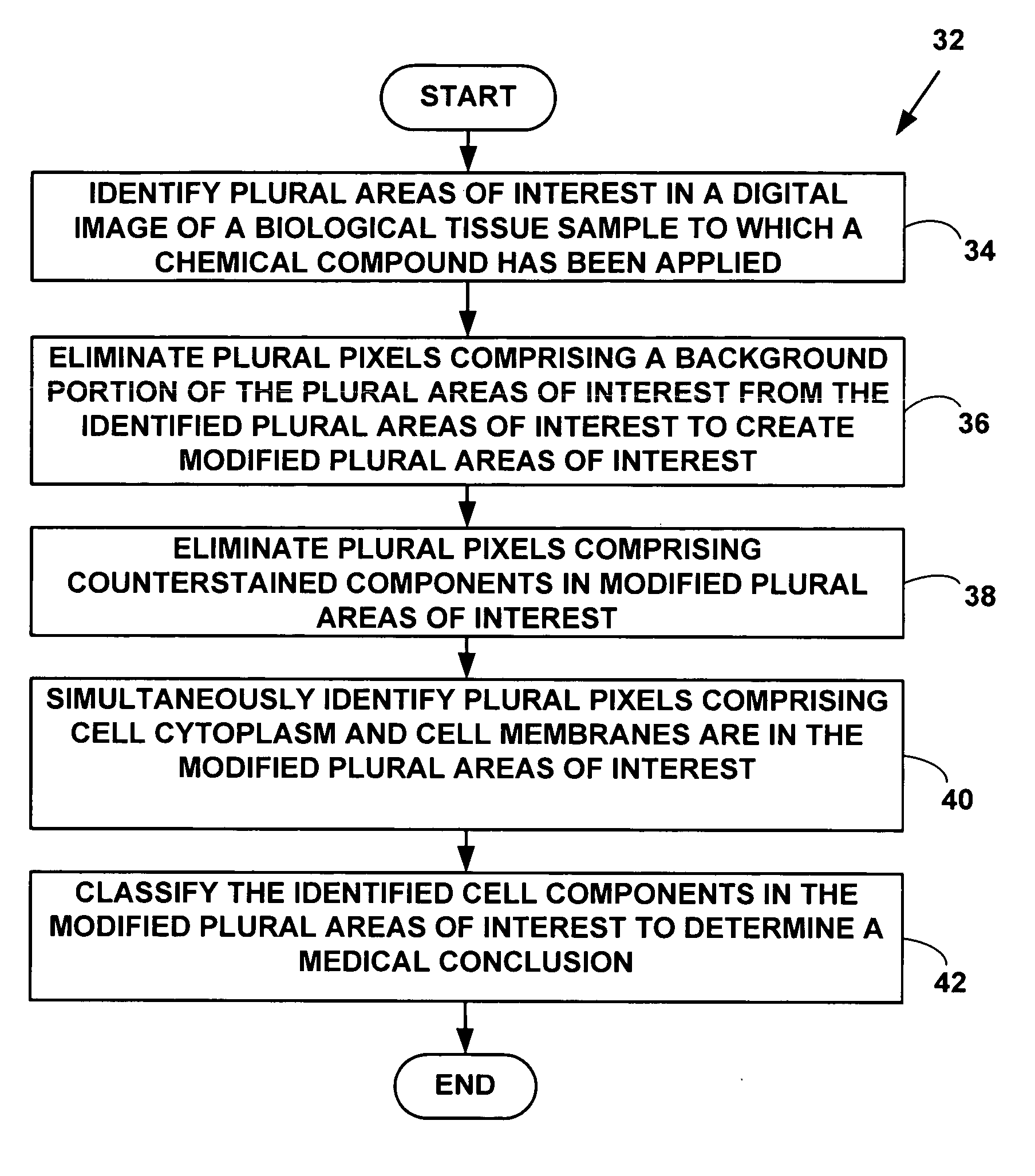
Limitation of the techniques available in prior art is the ability to identify and quantify individual cell components.
Each of the three components can get stained or unstained or counterstained.
Observing a three dimensional cell in two dimensions has its own issues.
Simultaneous identification of components becomes complex if more than one component behaves similar.
That is, if
nucleus and
cytoplasm take
stain and appear brown red, detecting the boundary between cytoplasm and membrane is much more difficult than the case where only membrane gets stained.
Similarly, it will be very difficult to locate
nucleus in a cell if both nucleus and cytoplasm are stained similar.
However, cocktail
staining is found to be unsuitable for some applications.
None of the existing products are capable of providing simultaneous quantitation of nucleus, cytoplasm and membrane, which is a key factor for studies of translocation.
However, these attempts still do not solve all of the problems with automated biological analysis systems that have been developed to improve the speed and accuracy of the testing process.

