
Biopsy Device With Variable Side Aperture

a biopsy device and variable side aperture technology, applied in medical science, surgery, vaccination/ovulation diagnostics, etc., can solve the problems of limiting the choice of access direction, difficult to biopsy lesions near the skin with a core biopsy probe, shortening the duration and inconvenience of the procedure, etc., to avoid discomfort and disfiguring scarring.

Inactive Publication Date: 2008-05-15
DEVICOR MEDICAL PROD
View PDF52 Cites 26 Cited by
  • Summary
  • Abstract
  • Description
  • Claims
  • Application Information

AI Technical Summary

Benefits of technology

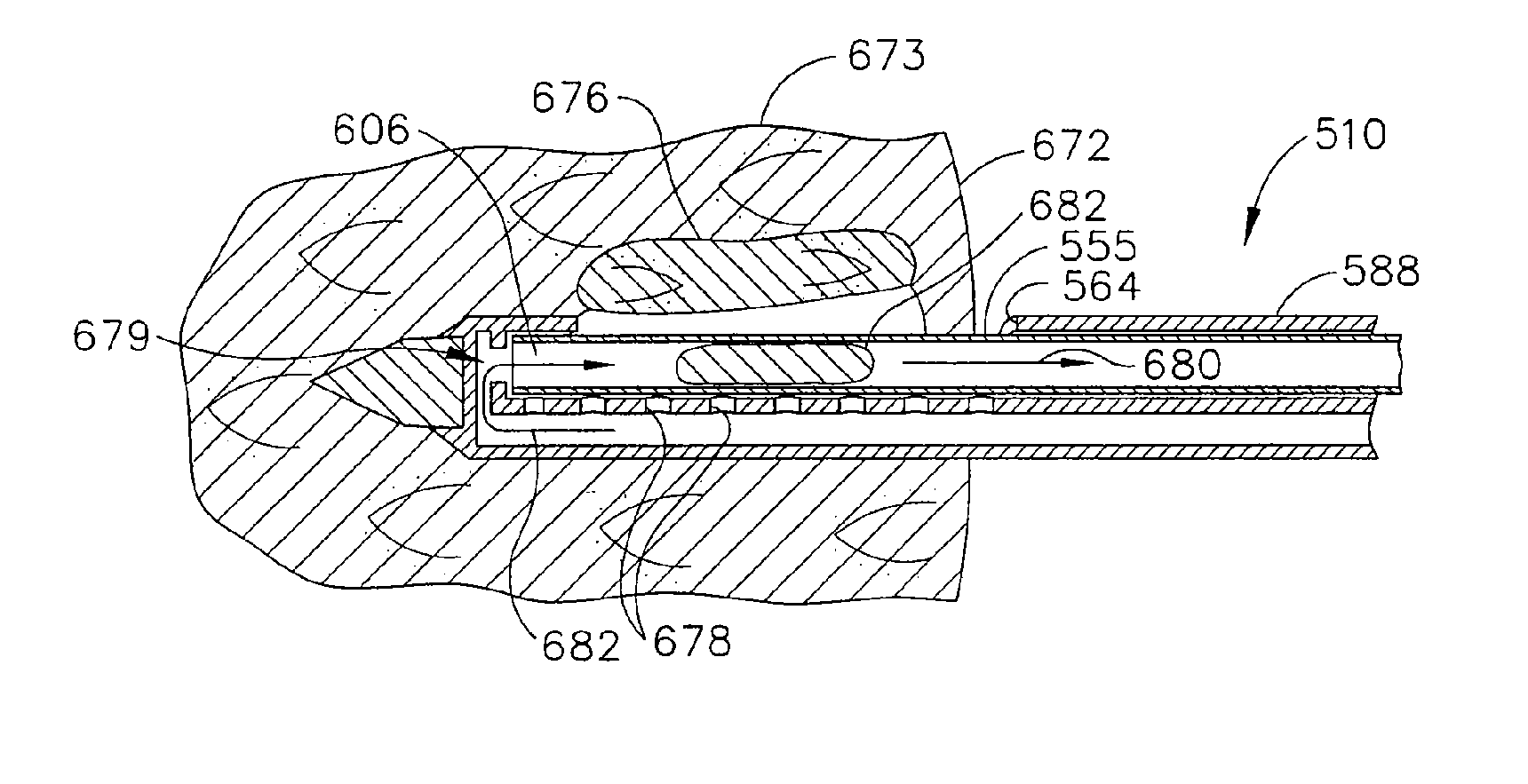
[0009] The present invention addresses these and other problems of the prior art by providing a core biopsy device having a probe assembly with a side aperture that is selectively longitudinally sized for taking samples. A proximal blocking member may be selectively positioned in the side aperture such that a proximal portion thereof is blocked when otherwise an outer layer of skin would prolapse into the side aperture when a cutter tube is retracted and then advanced to take a tissue sample. Thereby, discomfort and disfiguring scarring is avoided while still retaining the ability to take a tissue sample of a lesion near to a patient's skin.
[0010] In one aspect consistent with aspects of the invention, an apparatus for performing a core biopsy includes a curved portion sized to correspond to a portion of the probe surrounding at least the proximal portion of the side aperture which is held thereover by an engaging structure attached to the curved portion and registered to at least partially encompass and engage the probe. A gripping portion attached to the curved portion allows for a user to longitudinally position the curved portion over the proximal portion of the side aperture when desired. Thereby, an additional capability is provided for a biopsy device even when its operation requires that a cutter tube fully retract to remove a tissue sample before a subsequent translation for taking another sample.
[0011] In another aspect of the invention, a method of performing a biopsy of a lesion near to a patient's skin may be performed by translating a cutter tube distally in a cutter lumen to a distal-most position blocking a side aperture proximate to a distal end of the cutter lumen, inserting the cutter lumen through the patient's skin with the side aperture aligned with the lesion, retracting the cutter lumen to a position distal to an outer surface of the patient's skin with a proximal portion of the side aperture extending proximally thereof, prolapsing tissue into the side aperture, and translating the cutter lumen distally to cut a tissue sample. Using the cutter tube itself to block a portion of the side aperture that is exposed outside of the patient's skin avoids gouging the skin during cutting cycles.

Problems solved by technology

This shortens the duration and inconvenience of the procedure between taking samples.
However, there are situations in which lesions near the skin are difficult to biopsy with a core biopsy probe.
This is more often a challenge with a small breast, especially when compressed in a localization fixture that limits the choice in access direction.
If the side aperture of the probe is partially exposed, then vacuum assist may be ineffective as the specimen bowl in the probe is exposed to atmospheric pressure.
Further, skin may prolapse into the specimen bowl before the cutter advances into the tissue, causing gouging of the skin, increasing post-procedure pain and scarring.

Method used


Image

  • Biopsy Device With Variable Side Aperture
  • Biopsy Device With Variable Side Aperture
  • Biopsy Device With Variable Side Aperture

Examples


Embodiment Construction

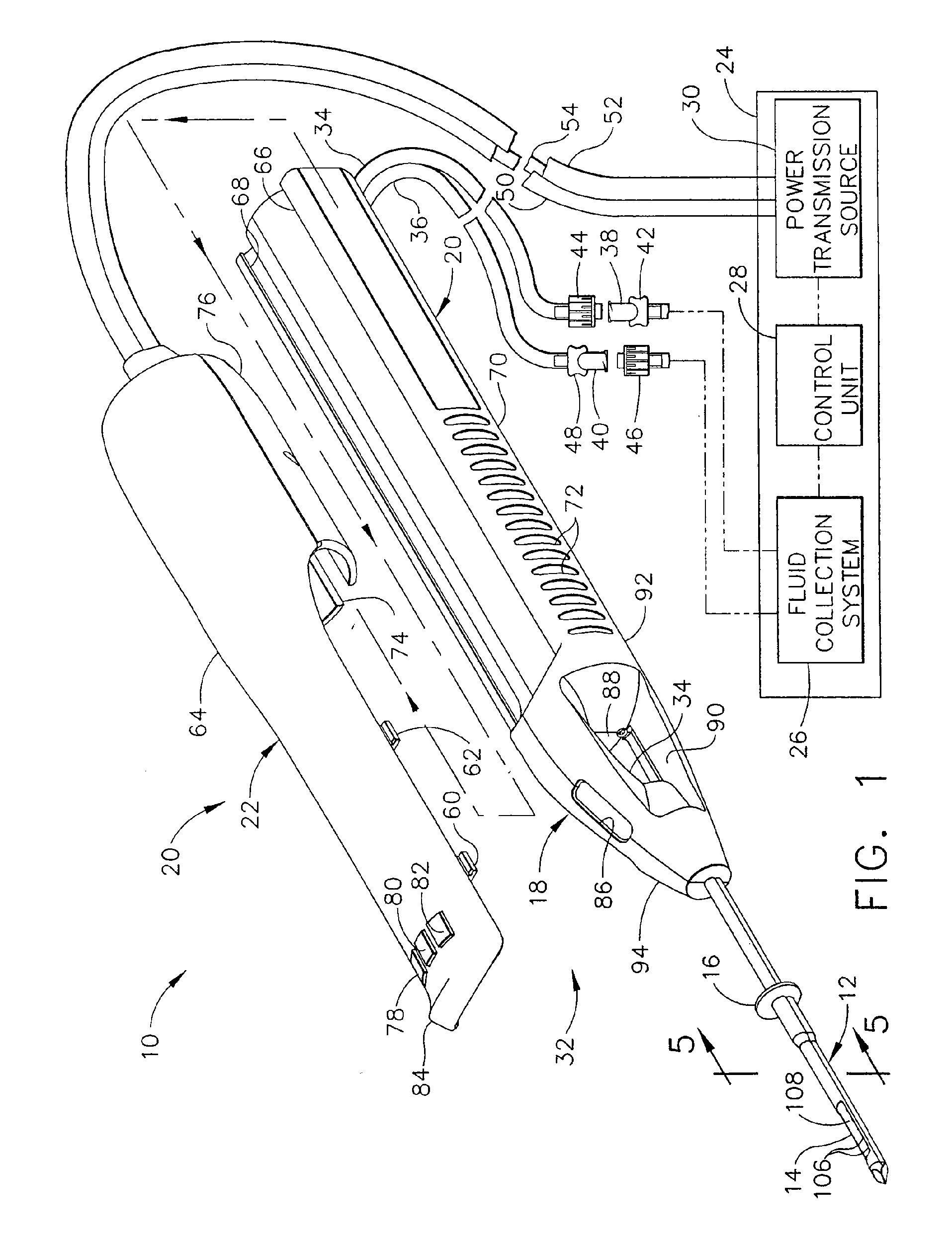
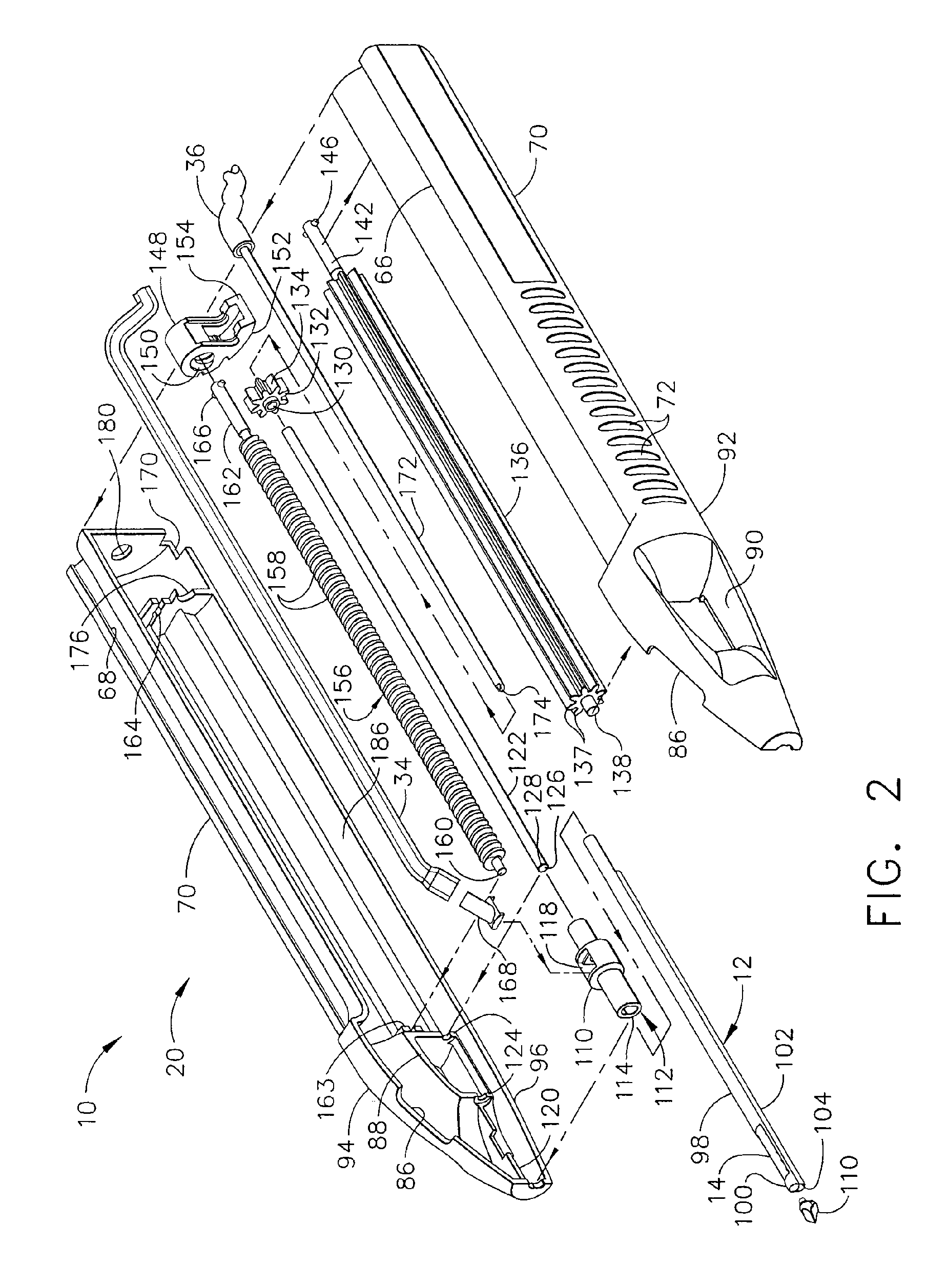
[0037] Core sampling biopsy devices are given additional flexibility to remove tissue samples that reside close to an insertion point by incorporating an ability to block a proximal portion of a side aperture in a probe, corresponding to where the outer tissue layers contact the probe when the distal portion of the side aperture is placed beside a suspicious lesion. This proximal blocking feature may be provided by a separate member attachable to generally-known biopsy devices, leveraging existing capital investments in an economical way. In the first illustrative version, a biopsy device that includes a long stroke cutter that retracts fully out of a probe between samples in order to retrieve tissue samples is thus adapted when a variable sized side aperture is desired. Alternatively, in a second illustrative version, a biopsy device that has tissue sample retrieval that is independent of cutter position is adapted to employ the cutter as the proximal blocking feature to achieve a ...


PUM


Abstract

A biopsy device and method are provided for obtaining a tissue sample, such as a breast tissue biopsy sample. The biopsy device may include an outer cannula having a distal piercing tip, a cutter lumen, a side tissue port communicating with the cutter lumen, and at least one fluid passageway disposed distally of the side tissue port. The inner cutter may be advanced in the cutter lumen past the side tissue port to sever a tissue sample. After the tissue sample is severed, and before the inner cutter is retracted proximally of the side tissue port, the cutter may be used to alternately cover and uncover the fluid passageway disposed distally of the side tissue.

Description

PRIORITY [0001] This application is a Divisional of prior, co-pending application Ser. No. 11 / 072,719, filed Mar. 4, 2005, which application is hereby incorporated by reference in its entirety.CROSS REFERENCE TO RELATED APPLICATIONS [0002] This application cross references and incorporates by reference the following commonly assigned patent applications: U.S. patent application Ser. No. 10 / 676,944, “Biopsy Instrument with Internal Specimen Collection Mechanism” filed Sep. 30, 2003 in the name of Hibner et al.; and U.S. patent application Ser. No. 10 / 732,843, “Biopsy Device with Sample Tube” filed Dec. 10, 2003 in the name of Cicenas et al. FIELD OF THE INVENTION [0003] The present invention relates in general to biopsy devices, and more particularly to biopsy devices having a cutter for severing tissue. BACKGROUND OF THE INVENTION [0004] The diagnosis and treatment of tissue is an ongoing area of investigation. Medical devices for obtaining tissue samples for subsequent sampling and...

Claims


Application Information

Patent Timeline
Patent Type & Authority Applications(United States)
IPC IPC(8): A61B1/00A61B10/00
CPCA61B10/0283A61B10/0275
Inventor WEIKEL, ROBERT F. JR.REICHEL, LEE E.HIBNER, JOHN A.LUDZACK, MICHAEL R.
Owner DEVICOR MEDICAL PROD
Learn More