Method of compensation of respiratory motion in cardiac imaging

Active Publication Date: 2010-12-09
SIEMENS HEALTHCARE GMBH
View PDF5 Cites 58 Cited by
  • Summary
  • Abstract
  • Description
  • Claims
  • Application Information

AI Technical Summary

Problems solved by technology

This alignment or registration of pre-operative volumetric image datasets with intra-operative images is a well-known technique to help overcome some of the drawbacks of X-ray imaging but it is a computationally demanding and challenging task.
Such re-registrations are challenging because of the low quality and lack of soft tissue contrast in intra-operative 2-D X-ray fluoroscopic images.
Further, re-registrations are also computationally expensive.
However, due to the relatively long breathing cycle of several seconds, the resulting imaging frequency of respiratory-gated angiograms is significantly reduced and is not practical for interventional applications.
Even if this method successfully reduces breathing motion by relatively simple and

Method used


Image

  • Method of compensation of respiratory motion in cardiac imaging
  • Method of compensation of respiratory motion in cardiac imaging
  • Method of compensation of respiratory motion in cardiac imaging

Examples


Example

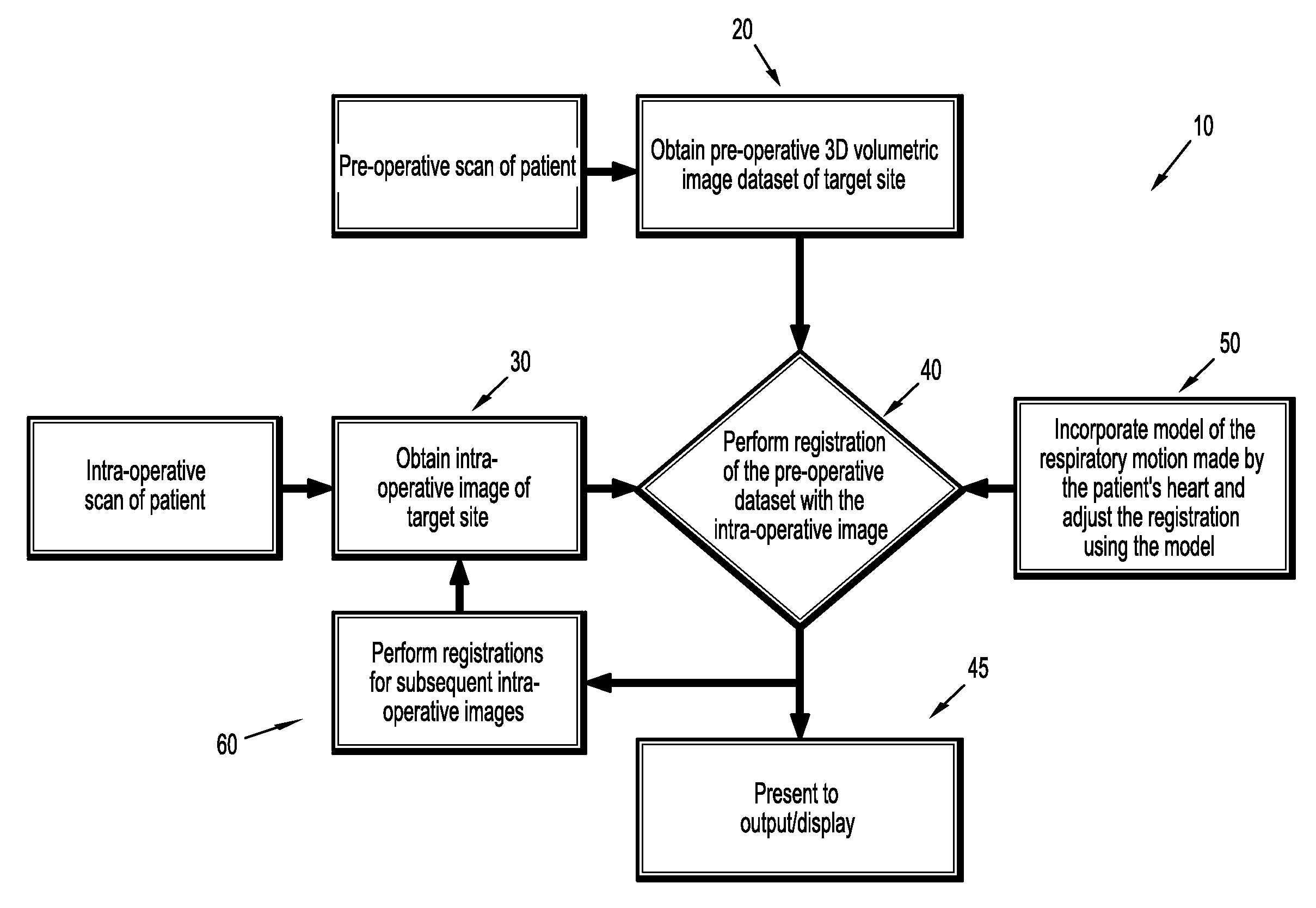
[0036]FIG. 1 shows an illustration of a method 10 for compensation of respiratory motion in cardiac imaging of a subject patient carried out in accordance with the present invention. Step 20 shows that at least one pre-operative volumetric image dataset of the target site of the patient is obtained. An example is a 3D coronary tree image around the target site which is reconstructed from a preparatory, pre-operative CT scan of the patient. The CT scan may be performed using any commercially available CT scanner. The volumetric image dataset may be stored in appropriate data storage and then obtained via retrieval from data storage as described below. As previously noted, the medical professional routinely arranges for pre-operative plans, like the pre-operative volumetric image dataset, to be integrated into the interventional surgery suite of tasks to provide an assist in complex non-invasive cardiac interventional procedures.

[0037]In step 30, at least one intra-operative image of ...


PUM


Abstract

A method (10) for respiratory motion compensation by applying principle component analysis (PCA) on cardiac imaging samples obtained using 2D/3D registration of a pre-operative 3D segmentation of the coronary arteries.

Description

CROSS-REFERENCE TO RELATED APPLICATIONS[0001]This application claims the benefit of Provisional U.S. Patent Application Ser. No. 61 / 185,340 entitled, “Model-Constrained Respiratory Motion Compensation for Cardiac Interventions”, filed in the name of Matthias Schneider, Hari Sundar, Rui Liao, and Chenyang Xu on Jun. 9, 2009, the disclosure of which is also hereby incorporated herein by reference.FIELD OF INVENTION[0002]The present invention relates to cardiac imaging. More particularly, the present invention relates to respiratory motion compensation in cardiac imaging.BACKGROUND OF THE INVENTION[0003]Complex non-invasive cardiac interventional procedures, such as, cardiac catheter ablation, carotid artery stenting, and percutaneous coronary interventions for chronic total occlusions, are routinely guided by X-ray coronary angiography that provides information about the motion and morphology of the coronary arteries under operation. Although it depends on the interventional procedure...

Claims


Application Information

Patent Timeline
IPC IPC(8): G06K9/00G06T15/00
CPCG06K9/6247G06K2209/05G06T7/0034G06T2207/30048G06T2207/10081G06T2207/10121G06T2207/20081G06T7/208G06T7/35G06T7/277G06V2201/03G06V10/7715G06F18/2135
Inventor SCHNEIDER, MATTHIASSUNDAR, HARILIAO, RUIXU, CHENYANG
Owner SIEMENS HEALTHCARE GMBH
Why Eureka
Try Eureka