Shape estimates and temporal registration of lesions and nodules

a technology of shape estimation and temporal registration, applied in image enhancement, image analysis, instruments, etc., can solve the problems of difficult detection of lung cancer, risk of spreading to lymph nodes in the lungs and mediastinum, and difficulty in lung cancer detection, so as to achieve accurate shape estimation of nodules

Active Publication Date: 2009-02-03
ICAD INC
View PDF3 Cites 10 Cited by
  • Summary
  • Abstract
  • Description
  • Claims
  • Application Information

AI Technical Summary

Benefits of technology

[0015]Accordingly, it is an object of the present invention to provide for a method for accurate shape estimation of nodules.

Problems solved by technology

Lung cancer is caused by abnormal cells growing within the lung tissue and has a risk of spreading to lymph nodes in the lungs and mediastinum.
Unfortunately, lung cancer is difficult to detect in its early stages.
A complicated anatomy combined with perceptual problems that accompany the projection of a three-dimensional object (the patient) into two dimensions (the image plane), however, makes identification of lung nodules a burdensome task for radiologists, resulting in a detection rate has been estimated to be between 20% and 50%, see Lorentz, et al., “Miss rate of lung cancer on the chest radiograph in clinical practice,”Chest, 115:720-724, 1999.
The detection of lung nodules in CT systems is still confounded by the presence of blood vessels in the lungs.
Although multiple slices provide more opportunities for a lesion to be detected, the possibility of missing a lesion is also increased due to the increased workload by generating a greater number of individual images per scan.

Method used


Image

  • Shape estimates and temporal registration of lesions and nodules
  • Shape estimates and temporal registration of lesions and nodules
  • Shape estimates and temporal registration of lesions and nodules

Examples


Embodiment Construction

[0035]In the following detailed description of the preferred embodiments, reference is made to the accompanying drawings that form a part hereof, and in which are shown by way of illustration, and not by way of limitation, specific preferred embodiments in which the invention may be practiced. It is to be understood that other embodiments may be utilized and that logical, mechanical and electrical changes may be made without departing from the spirit and scope of the present invention.

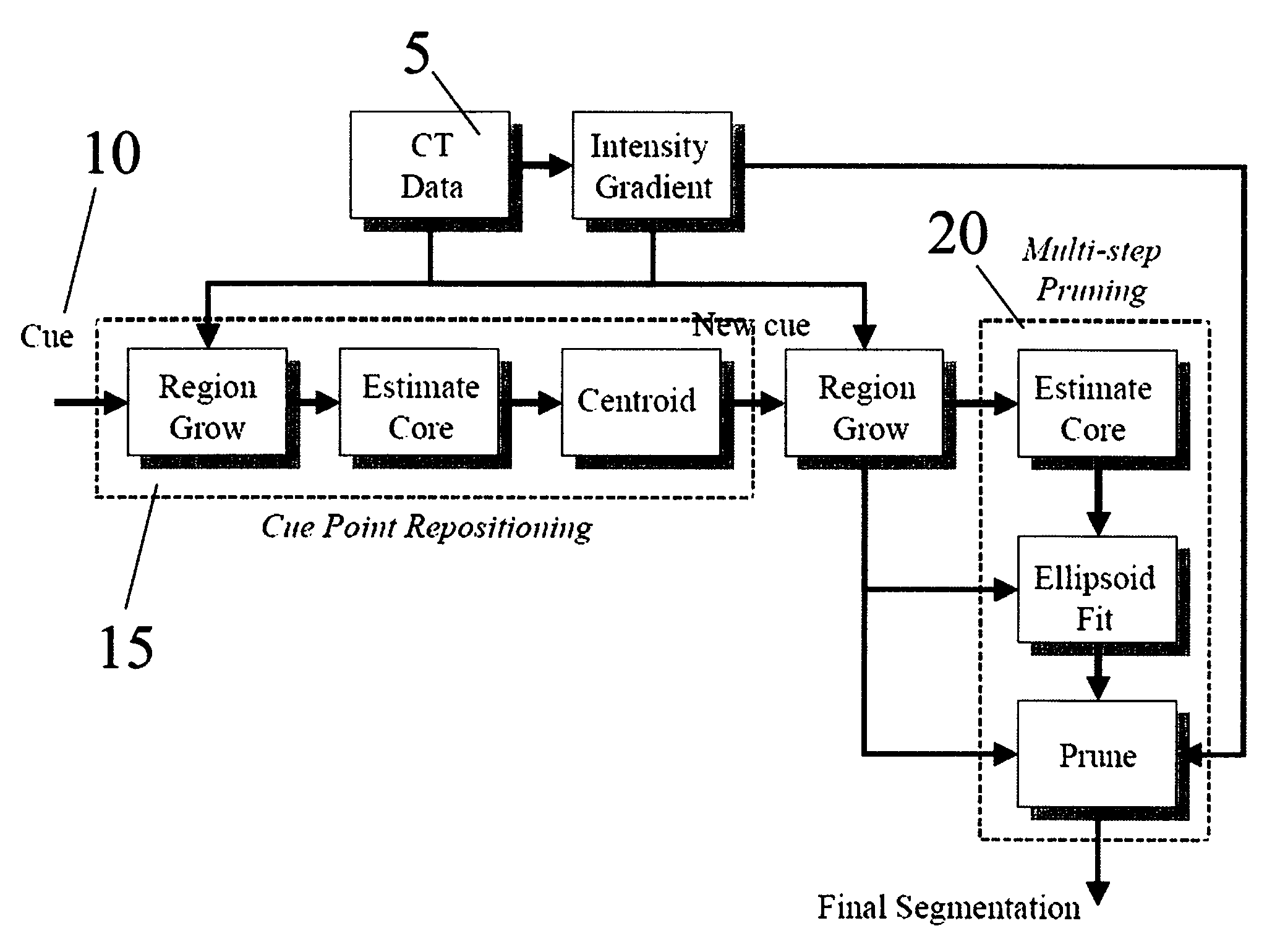
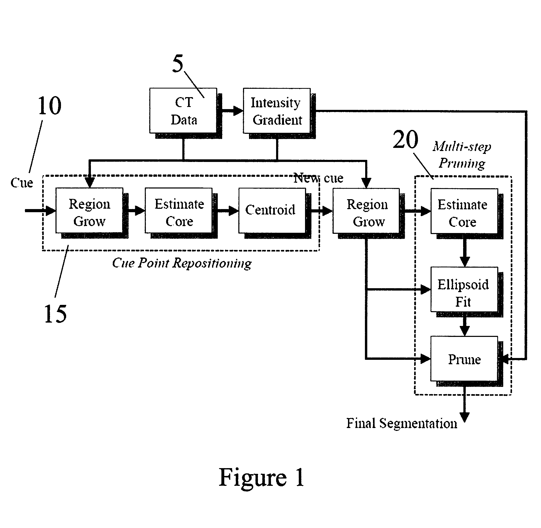
[0036]A CAD and analysis system can help physicians quickly and accurately identify and analyze problematic regions of the body. For example, CAD systems are used to detect pulmonary nodules and colonic polyps using CT imagery. This invention addresses one part of such as system, a tool designed to segment regions with minimal cueing from radiologists. In a CAD driven embodiment, cueing is provided by detection algorithms. In an interactive embodiment, cues are provided by a user who indicates a cue po...


PUM


Abstract

A method for refining shape estimates of detected abnormalities in medical images, particularly for the detection of pulmonary lesions in CT imagery is described. A cue point is refined prior to an initially segmentation of the lesion. A radial gradient is computed for points on an initial segmented lesion. Portions of the lesion with radial gradients deviating beyond a threshold angle are removed from the lesion. Registering imagery from more than one CT exam uses a high intensity structure, essentially bone, to provide coarse and fine alignment of a set of two-dimensional images is also described. A MIP image is formed from the three-dimensional images. A second MIP image is then formed from imagery. The second MIP image is correlated with the first MIP image to determine a preferred registration location.

Description

CROSS-REFERENCE TO RELATED APPLICATIONS[0001]This application claims the benefit of U.S. Provisional Application No. 60 / 524,837, filed Nov. 25, 2003, incorporated herein by reference.BACKGROUND OF THE INVENTION[0002]1. Field of the Invention[0003]The present invention generally relates to computer-aided detection (CAD) of suspicious regions in three-dimensional medical imagery and, in particular, relates to improving shape estimates of nodules detected in three-dimensional imagery of lungs as well as registering three-dimensional imagery from multiple computed tomography (CT) exams.[0004]2. Discussion of Background[0005]Lung cancer is now the most common form of cancer diagnosed in the United States and remains a leading cause of cancer death in the United States in both males and females. Lung cancer accounts for 14% of all cancer diagnoses and over 30% of all cancer deaths. The American Cancer Society estimates approximately 173,000 new lung cancer cases will be detected and some ...

Claims


Application Information

Patent Timeline
Patent Type & Authority Patents(United States)
IPC IPC(8): G06K9/00G06T5/00G06T7/00
CPCG06T7/0012G06T7/0028G06T7/0038G06T7/0081G06T7/0089G06T2207/10081G06T2207/20116G06T2207/20156G06T2207/30064G06T7/33G06T7/38G06T7/11G06T7/149
Inventor GURCAN, METIN N.HARDIE, RUSSELL C.ROGERS, STEVEN K.
Owner ICAD INC
Why Eureka
Try Eureka