Living small animal imaging system and imaging method

An imaging system and small animal technology, applied in the field of biomedical imaging, can solve the problems of erroneous diagnosis results, inability to locate molecular information, and inability to provide sample structure information, and achieve the effect of improving accuracy.

Inactive Publication Date: 2010-02-24
HUAZHONG UNIV OF SCI & TECH
View PDF2 Cites 40 Cited by
  • Summary
  • Abstract
  • Description
  • Claims
  • Application Information

AI Technical Summary

Problems solved by technology

Although fluorescence tomography can reconstruct fluorescent molecular markers in small animals, it cannot image the structure of small animal samples
Therefore, the molecular information obtained by pure fluorescence tomography cannot be accurately located in small animals.
This can easily lead to wrong diagnostic results (Ralf B. Schulz et al., Comparison of noncontact and fiber-based fluorescence-mediated tomography, "Optics Letters", 2006: 769-771)
Chinese Patent Publication No. CN 101057788A, published on October 24, 2007, the name of the invention is a fluorescent molecular tomography imagi

Method used


Image

  • Living small animal imaging system and imaging method
  • Living small animal imaging system and imaging method

Examples


Embodiment Construction

[0025] specific implementation plan

[0026] In order to make the object, technical solution and advantages of the present invention clearer, the implementation manner of the present invention will be further described in detail below in conjunction with the accompanying drawings.

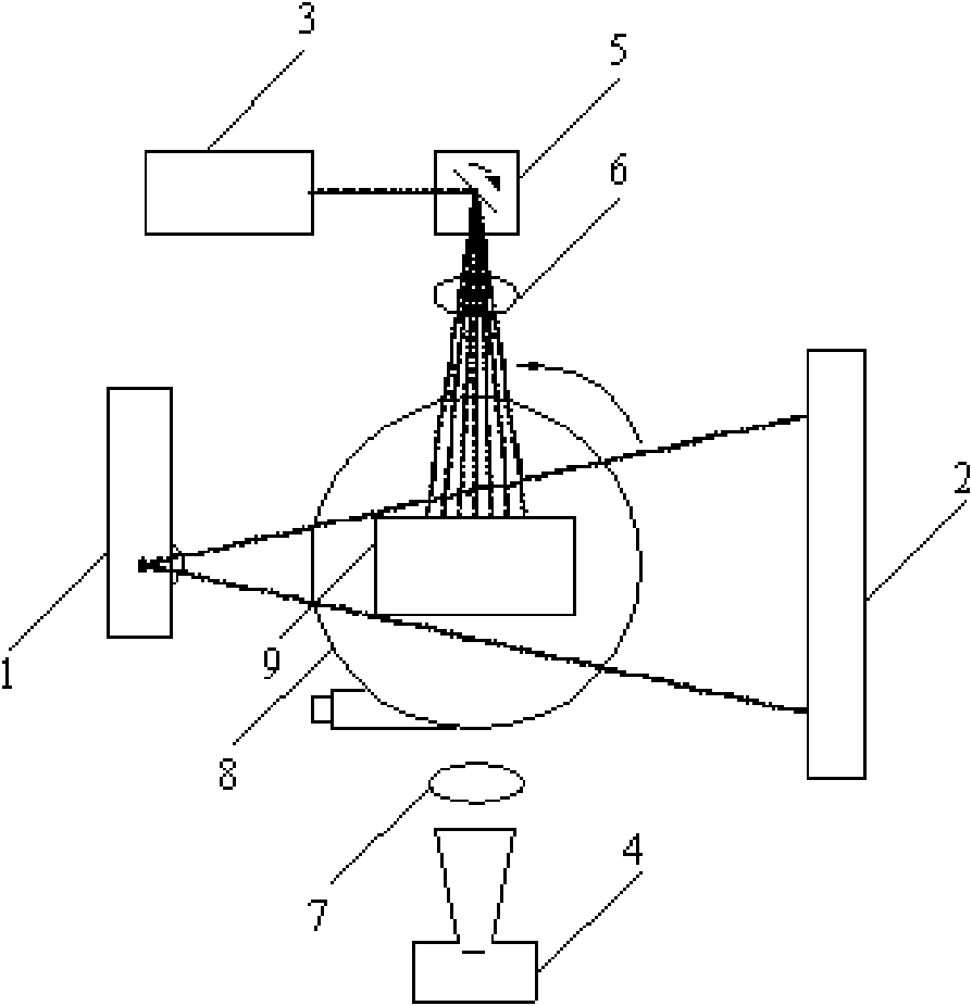
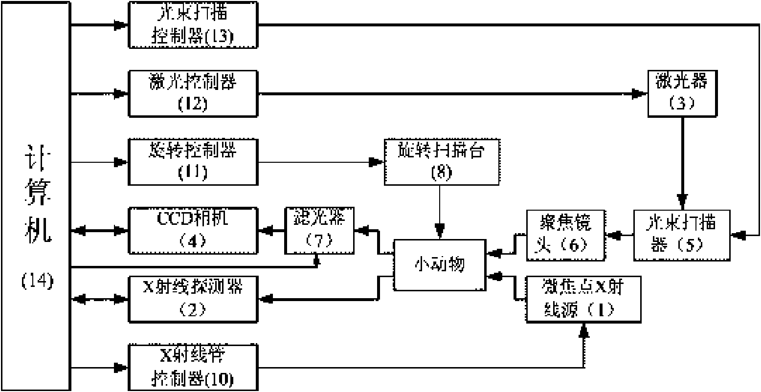
[0027] Such as figure 1 , figure 2 As shown, the present invention belongs to a dual-mode imaging system, which includes a fluorescence tomography subsystem and a micro-CT subsystem, and couples the two imaging modes into one system by design to realize dual-mode imaging of small animals in vivo . The imaging system includes a main control computer 14, a ray source and an X-ray detection part, an excitation light source and an excitation light / fluorescence detection part, and a rotating scanning part.

[0028] The micro-focus ray source 1 and the X-ray detector 2 are respectively placed on both sides of the sample, and X-rays pass through the sample and are projected on the imaging surface of t...


PUM

PropertyMeasurementUnit
Wavelengthaaaaaaaaaa
Thicknessaaaaaaaaaa
The inside diameter ofaaaaaaaaaa

Abstract

The invention discloses a living small animal imaging system in a miniature CT and fluorescent tomography dual mode and an imaging method thereof. The living small animal imaging system in the miniature CT and the fluorescent tomography dual mode comprises a main control computer, an X-ray source and an X-ray detection device, an excitation source and an excitation light/fluorescent light detection device, and a rotary scanning device, wherein the X-ray source and the X-ray detection device, the excitation source and the excitation light/fluorescent light detection device, and the rotary scanning device are controlled by the main control computer. The invention can simultaneously acquire structure information by carrying out the miniature CT imaging on a small animal and fluorescent-labeled molecular information by carrying out the fluorescent tomography on small animals to enable the molecular information acquired by the fluorescent tomography to be accurately positioned in the smallanimal, thereby being beneficial to improving the diagnosis accuracy.

Description

technical field [0001] The invention relates to the technical field of biomedical imaging, in particular to an imaging system and imaging method for living small animals. Background technique [0002] Molecular imaging is an imaging method emerging in recent years that can dynamically observe biological events at the cellular and molecular levels in small living animals. It can be applied to early diagnosis of cancer, dynamic detection of tumor growth and metastasis, development of new drugs, etc. Mainly use traditional medical instruments such as MRI (magnetic resonance imaging, magnetic resonance imaging), PET (positron emission tomography, positron emission tomography), SPECT (single photonemission computed tomography, single photon emission tomography), etc., combined with different Molecular probes for molecular imaging. Fluorescence tomography is a non-destructive optical tomography technology. Compared with traditional imaging methods, its advantages are that it is ...

Claims


Application Information

Patent Timeline
IPC IPC(8): A61B5/00A61B6/03
CPCA61B6/508
Inventor 骆清铭龚辉杨孝全全国涛邓勇
Owner HUAZHONG UNIV OF SCI & TECH
Why Eureka
Try Eureka