Structured light quick scanning microscopic imaging method

A fast scanning and microscopic imaging technology, applied in microscopes, optics, optical components, etc., can solve the problems of poor system reliability, redundant data volume, slow imaging speed, etc., to save sample movement time, improve imaging speed, reduce Modulation of velocity-dependent effects

Active Publication Date: 2013-07-17
WUHAN OE BIO CO LTD
View PDF4 Cites 25 Cited by
  • Summary
  • Abstract
  • Description
  • Claims
  • Application Information

AI Technical Summary

Problems solved by technology

[0008] The main purpose of the present invention is to overcome the problems of slow imaging speed, poor system reliability and excessive redundant data volume when the exi

Method used


Image

  • Structured light quick scanning microscopic imaging method
  • Structured light quick scanning microscopic imaging method
  • Structured light quick scanning microscopic imaging method

Examples


Example Embodiment

[0025] The specific embodiments of the present invention will be described below with reference to the drawings.

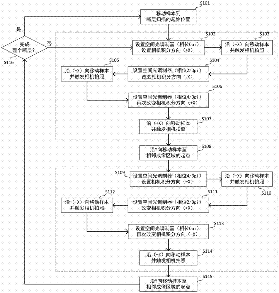
[0026] figure 1 It is a flowchart of the present invention. The flow chart shows a complete tomographic scanning process, which takes a total of 6 linear scanning processes (S103, S105, S107, S110, S112, and S114) of 2 imaging positions as a scanning cycle, and executes this cycle repeatedly until the tomography The scan is complete. For example: at the end of step S102 to set the spatial light modulator phase 0pi and set the camera integration direction (+X), the sample is in a static state, and step S103 the sample starts to move along the +X direction and triggers the camera to take a photo. After completion, the sample is in a static state Step S104 sets the spatial light modulator phase 2 / 3pi, and changes the camera integration direction -X, step S105 the sample starts to move along the -X direction and triggers the camera to take pictures, after completion the...


PUM


Abstract

The invention relates to a structured light quick scanning microscopic imaging method. The method comprises the following steps of: (1) controlling a spatial light modulator to generate a modulation pattern with single spatial frequency by a computer, and projecting the modulation pattern on a sample fracture; (2) under the condition that the phase of the modulation pattern is unchanged, driving a sample to move by an electric control movement platform, and synchronously triggering a line-scan digital camera to take a picture; (3) changing the phase of the modulation pattern, and scanning the same imaging region until different lighting phase images required by structured light imaging is acquired; and (4) moving a sample, and executing the steps (2) to (4) for the subsequent adjacent imaging region until the whole sample fracture is scanned. By performing phase change for one time before scanning motion is performed at each time, requirements of imaging on the speed and robustness of the modulator are reduced; and in single-time scanning motion, a complete image is acquired, and adjacent images are partially overlapped. Sample motion and camera exposure are synchronously performed, so that time is saved, and imaging speed is increased.

Description

technical field [0001] The invention relates to a microscope imaging method, in particular to a large-sample fast imaging method based on a structured light microscope. Background technique [0002] A microscope is a precision optical instrument with a history of more than 300 years. It uses optical principles to magnify and image tiny objects that cannot be distinguished by the human eye. With the aid of microscopes, people have seen many tiny organisms and the basic unit of organisms—cells—that were invisible in the past. The various performances of the microscope, especially its resolution, are related to the ability of human beings to understand the microscopic world. According to Rayleigh's criterion, conventional optical microscopes have resolution limits in both lateral and axial directions. But in contrast, the axial resolution is often only a few microns to tens of microns, which affects the tomographic ability of traditional optical microscopes. [0003] Mechani...

Claims


Application Information

Patent Timeline
IPC IPC(8): G02B21/36
Inventor 骆清铭龚辉许冬力李安安
Owner WUHAN OE BIO CO LTD
Why Eureka
Try Eureka