Viral vector production system

A technology for infectious diseases, detection reagents, used in the field of biomarker detection and treatment of infection and infectious disease conditions

Active Publication Date: 2016-10-12
MOREHOUSE SCHOOL OF MEDICINE
View PDF3 Cites 6 Cited by
  • Summary
  • Abstract
  • Description
  • Claims
  • Application Information

AI Technical Summary

Problems solved by technology

Continuous HBV replication increases the risk of cirrhosis and HCC progression

Method used


Image

  • Viral vector production system
  • Viral vector production system

Examples


Embodiment 1

[0148] Example 1: Materials and methods

[0149] patient

[0150] Patients with hepatitis were recruited for this study from four clinical sites in the Atlanta metropolitan area. All samples were collected according to protocols approved by the Institutional Review Board and Human Trials Research Committee at Mauhurs School of Medicine, and informed consent was obtained from all patients and healthy volunteers according to the guidelines established by the Institutional Review Board. Relevant information is also collected from the patient's medical records.

[0151] Sample Collection and Storage

[0152] Urine samples were collected from routine patient visits. Obtain clinical data from patient medical records. Urine is collected in a sterile container and shipped back to the laboratory. Urinalysis was performed on each sample using Multistix 10SG reagent strips (Bayer, Elkhart, IN) and albumin-to-creatine ratio determined by Siemens Clinitek microalbumin test strips (B...

Embodiment 2

[0173] Example 2: Sandwich ELISA of Hepatitis B Virus Biomarkers in Urinary Exosomes from Patients analyze

[0174] Urine exosomes isolated from hepatitis patients and normal control patients were analyzed for hepatitis A virus (HAV), hepatitis B virus (HBV), and hepatitis C virus (HCV)-related organisms by exosome ELISA presence of markers.

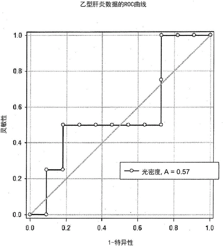
[0175] figure 1 Receptor action characteristic (ROC) curves for detection of hepatitis B using anti-hepatitis B antibodies in an exosome ELISA are shown.

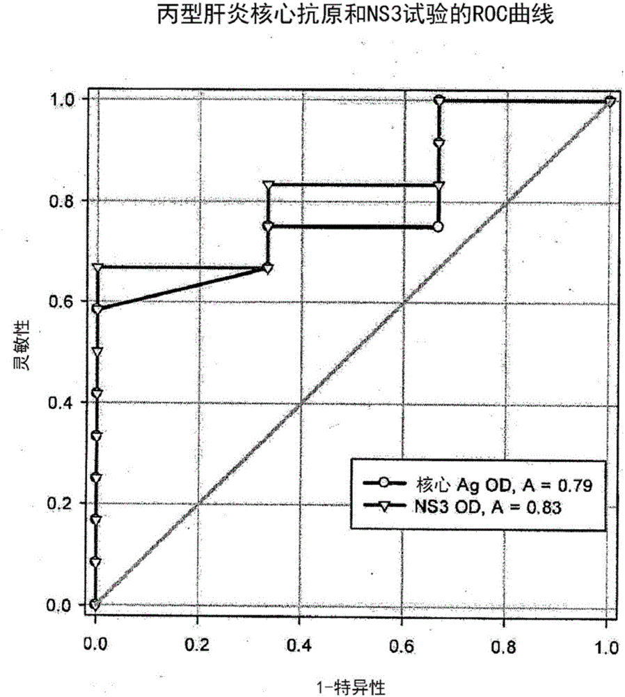
[0176] figure 2 is the ROC curve for the detection of hepatitis C in an exosome ELISA using HCV core antigen and HCV NS3 protein as biomarkers for hepatitis C.


PUM

PropertyMeasurementUnit
diameteraaaaaaaaaa

Abstract

The present invention relates to a nucleic acid sequence comprising a binding site operably linked to a nucleotide of interest, wherein the binding site is capable of interacting with an RNA-binding protein such that translation of the nucleotide of interest is repressed in a viral vector production cell.

Description

[0001] This application claims priority to US Provisional Application Serial No. 61 / 929,570, filed January 21, 2014. The entire contents of the aforementioned applications are incorporated herein by reference. technical field [0002] The present application relates generally to methods for detection and treatment, and in particular, to methods for detection and treatment of infection and infectious disease conditions targeting biomarkers present in exosomes secreted by bodily fluids. Background technique [0003] Exosomes are small vesicular bodies with a diameter of 40-100nm, which are secreted by many different cell types and communicate with other cells through the proteins and RNA they carry. Exosomes are generated intracellularly when cell membrane fragments spontaneously invaginate and are phagocytosed. The internalized fragments are broken into small vesicle bodies, which are subsequently expelled from the cell. The latter stage occurs when secondary endosomes, whi...

Claims


Application Information

Patent Timeline
Patent Type & Authority Applications(China)
IPC IPC(8): C12Q1/02C12Q1/68G01N33/92G01N33/569
CPCC12Q1/706C12Q2600/178G01N33/56994G01N2800/52
Inventor 加莱·W·纽曼萨姆·阿尼安维
Owner MOREHOUSE SCHOOL OF MEDICINE
Why Eureka
Try Eureka