System and method for single-exposure digital subtraction angiography imaging

An angiography and digital subtraction technology, applied in the field of medical X-ray imaging systems, can solve the problems of inaccurate matching of mask images and angiography images, differences and overlaps of energy spectra, and reduced quality of subtraction images, so as to improve diagnosis. Efficiency and equipment utilization, avoiding motion artifacts, reducing effects of radiation damage

Active Publication Date: 2017-04-19
NANJING UNIV OF AERONAUTICS & ASTRONAUTICS
View PDF4 Cites 10 Cited by
  • Summary
  • Abstract
  • Description
  • Claims
  • Application Information

AI Technical Summary

Problems solved by technology

Its disadvantage is that due to the voluntary or involuntary movement of the patient during the photography process, the mask image and the angiographic image cannot be accurately matched, resulting in poor registration artifacts or blu

Method used


Image

  • System and method for single-exposure digital subtraction angiography imaging

Examples


Embodiment 1

[0028] Example 1: Cardiovascular DSA Imaging

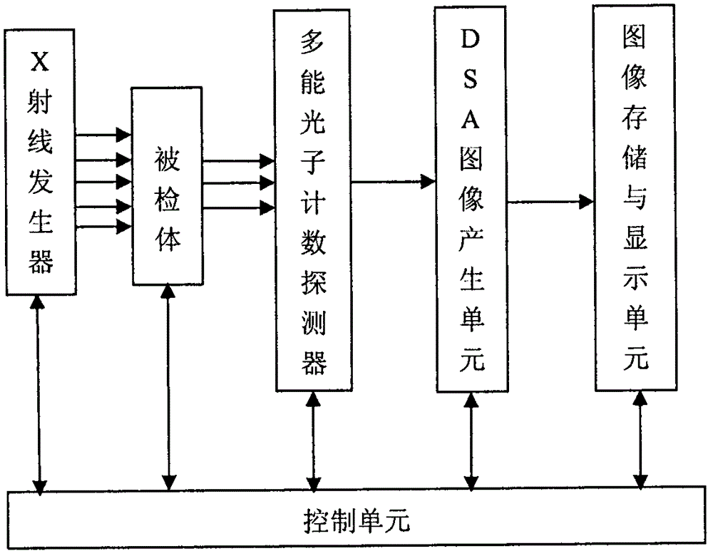
[0029] (1) Place the subject on the examination table, adjust its posture, and adjust the posture of the X-ray generator and detector to suit the DSA imaging of the cardiovascular system of the examined part;

[0030] (2) Set the energy threshold of the photon counting detector according to the properties of the contrast agent, and the weights α and β of the high and low energy segment images;

[0031] (3) Under the control of the control unit, start the x-ray generator to generate x-rays to pass through the cardiovascular part of the subject, and the transmitted x-rays are accepted by the multi-energy photon counting detector and counted in energy segments; DSA image generation The unit collects the signal output by the multi-energy photon counting detector, and after logarithmic processing, the image I of the high and low energy intervals is obtained H and I L , and apply the corresponding weighting coefficients α and β, and p...


PUM


Abstract

The invention discloses a system and method for single-exposure digital subtraction angiography (DSA) imaging. The system is composed of an x-ray generator, a multi-energy photon counting detector, a DSA image generation unit, a device for image storage and display, a sickbed, a rotary rack and a controller, wherein the x-ray generator can generate continuous energy spectrum x rays; the multi-energy photon counting detector detects x photons which penetrate a human body and can judge and count energy of the x photons respectively; the DSA image generation unit collects output signals of the multi-energy photon counting detector to form data of two transmission images of a high energy zone and a low energy zone; and DSA images are obtained through subtraction computation of the data, and a conventional x-ray image can be obtained through summary computation. The DSA image can be obtained by the system through single exposure, so motion artifacts can be avoided; the contrast medium dosage and the radiation agent dosage can be reduced; equipment efficiency can be increased; more effective approaches can be provided for DSA imaging of dynamic objects; and the system has important values in clinical application.

Description

[0001] Technical field: [0002] The present invention relates to a medical X-ray imaging system and method, specifically to a single-exposure digital subtraction angiography (DSA) imaging system and method, providing an effective means of low-dose imaging for the diagnosis and treatment of vascular lesions. [0003] Background technique: [0004] Cardiovascular disease is one of the leading causes of death worldwide, and obtaining accurate images of cardiovascular patients is of great significance for diagnosis and treatment. Angiographic imaging (DSA) is a large-scale real-time imaging diagnostic equipment, especially in coronary angiographic diagnosis, etc. It is an essential equipment, and it is also a platform for interventional therapy. It is mainly used for coronary stent implantation, minimally invasive valve implantation, interventional therapy for tumors, interventional therapy for cerebrovascular malformations and aneurysms, interventional therapy for uterine fibroid...

Claims


Application Information

Patent Timeline
IPC IPC(8): A61B6/00
CPCA61B6/4411A61B6/481A61B6/504A61B6/5211
Inventor 周正东
Owner NANJING UNIV OF AERONAUTICS & ASTRONAUTICS
Why Eureka
Try Eureka