Separating probe combination for diagnosing MITF (Microphthalmia-associated Transcription Factor) translocation kidney cancer and application of separating probe combination

A technology for probes and kidney cancer, applied in the field of fluorescent in situ hybridization probes, can solve the problem of not being able to screen out MITF translocation tumors, achieve a reliable success rate, and improve the effect of accuracy

Active Publication Date: 2017-10-27
NANJING GENERAL HOSPITAL NANJING MILLITARY COMMAND P L A
View PDF4 Cites 4 Cited by
  • Summary
  • Abstract
  • Description
  • Claims
  • Application Information

AI Technical Summary

Problems solved by technology

The two designs have their own advantages and disadvantages, and the detection results of the fusion probe are more accurate and specific. However, because MITF gene translocation tumors have not been discovered in the past, only our resear

Method used


Image

  • Separating probe combination for diagnosing MITF (Microphthalmia-associated Transcription Factor) translocation kidney cancer and application of separating probe combination
  • Separating probe combination for diagnosing MITF (Microphthalmia-associated Transcription Factor) translocation kidney cancer and application of separating probe combination
  • Separating probe combination for diagnosing MITF (Microphthalmia-associated Transcription Factor) translocation kidney cancer and application of separating probe combination

Examples


Embodiment 1

[0035] Embodiment 1: the preparation of DNA probe combination:

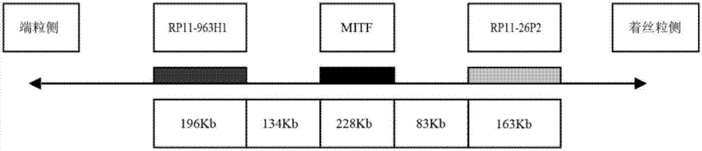
[0036] Select two BAC clone fragments that can be connected at both ends of the MITF gene on chromosome 3, control the farthest distance between the probes at both ends within 1500kb, keep a certain distance between the BAC clone fragments without overlapping, and the fragment sizes are similar. The cloned fragments were obtained from the Human BAC Cloning Center of EmpireGenomics (http: / / www.empiregenomics.com / helixhq / clonecentral / search / human). The centromeric BAC clone fragment of MITF was RP11-26P2 (fragment length 163kb), and the BAC clone fragment of MITF telomeric side was RP11-963H1 (fragment length 196kb). The sequence of linkage between the BAC clone fragment and the MITF gene is RP11-26P2, MITF, RP11-963H1. The positioning structure of the probe assembly is as follows figure 1 shown. Use the gap translation method to mark the BAC clone fragment on the telomere side with fluorescence of any color, pr...

Embodiment 2

[0038] Embodiment 2: fluorescence in situ hybridization process:

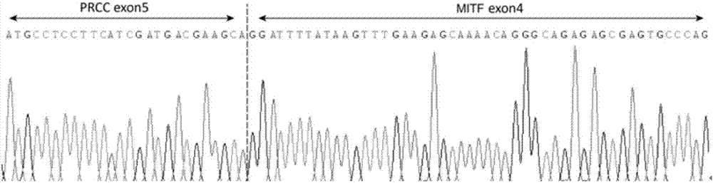
[0039] A case of MITF translocation renal carcinoma was diagnosed by high-throughput sequencing and RT-PCR detection of fusion genes ( figure 2 , corresponding to the results of this experiment can better reflect the reliability of this experiment), and two experienced pathologists referred to the WHO2016 urinary and male reproductive system classification standards, collected 30 cases of renal cell carcinoma diagnosed in Nanjing General Hospital of Nanjing Military Region as control group.

[0040] Wax blocks were sliced ​​at a thickness of 3 μm. After dewaxing, they were placed in 100%, 85%, and 70% ethanol for 2 minutes each, and then immersed in deionized water for 15 minutes in a water bath at 100°C. Put the tissue slices into pepsin K solution (0.1g pepsin, 40ml 0.01M HCL) at 37°C for 15min; rinse twice with 2×SSC (sodium chloride, sodium citrate) for 5min each, and place the slices in 0.1mol / L Soak in...


PUM


Abstract

The invention discloses a separating probe combination for diagnosing MITF (Microphthalmia-associated Transcription Factor) translocation kidney cancer and application of the separating probe combination. The separating probe combination for diagnosing the MITF translocation kidney cancer consists of a BAC (Bacterial Antigen Complex) cloning probe RP11-26P2 and a BAC cloning probe RP11-963H1. According to the separating probe combination disclosed by the invention, a fluorescence labeled DNA (Deoxyribonucleic Acid) combination combined at two ends of an MITF gene is designed according to the characteristics of the MITF translocation kidney cancer; in-situ hybridization, detection fusion and signal separation are carried out on the basis of paraffin embedded tissue sections, so that the accuracy rate of diagnosing tumors can be greatly increased. The probe combination for detecting the MITF translocation kidney cancer, provided by the invention, has the advantages of convenience,` quickness, reliability and high success rate, can be used for preparing a diagnosis kit for the MITF translocation kidney cancer, and provides a novel tool for quickly and accurately diagnosing the MITF translocation kidney cancer.

Description

technical field [0001] The invention belongs to the application field of fluorescence in situ hybridization probes, and relates to a separation probe combination for diagnosing MITF translocation renal cancer and its application in preparing MITF translocation renal cancer diagnostic reagents. Background technique [0002] In 2016, a new type of renal cell carcinoma was added to the new WHO classification of kidney tumors by histopathology: MiT family translocation renal cell carcinoma. The MiT family is the abbreviation of the microphthalmia-associated transcription factor family, which includes MITF, TFE3, TFEB and TFEC genes. [0003] MiT family translocation renal cell carcinomas that have been discovered so far include Xp11.2 translocation / TFE3 gene fusion-related renal cell carcinoma and t(6;11)(p21;q12) translocation / TFEB gene fusion-related renal cell carcinoma. Tumors with MITF and TFEC gene translocations have not been reported in the literature worldwide. [000...

Claims


Application Information

Patent Timeline
IPC IPC(8): C12Q1/68C12N15/11
CPCC12Q1/6841C12Q1/6886C12Q2600/166C12Q2563/107
Inventor 饶秋夏秋媛魏雪王璇李芳秋周晓军
Owner NANJING GENERAL HOSPITAL NANJING MILLITARY COMMAND P L A
Why Eureka
Try Eureka