Near-infrared two-zone confocal microscopic imaging system based on multidimensional adjustment frame

A confocal microscopy, multi-dimensional adjustment technology, used in medical science, analysis using fluorescence emission, catheters, etc., can solve the problem of less imaging equipment, and achieve high response rate, large penetration depth, and high signal-to-noise ratio. Effect

Active Publication Date: 2020-04-07
ZHEJIANG UNIV
View PDF14 Cites 2 Cited by
  • Summary
  • Abstract
  • Description
  • Claims
  • Application Information

AI Technical Summary

Problems solved by technology

[0003] Due to the limitations of imaging devices and fluorescent probes, the excitation light and emitted fluorescence signals used by confocal scanning microscopes are often in the visible light (400~760 nm) or near-infrared region 1 (760~900 nm) band, based on the near-infrared region 2 (900~1700 nm) band imaging equipment is less

Method used


Image

  • Near-infrared two-zone confocal microscopic imaging system based on multidimensional adjustment frame
  • Near-infrared two-zone confocal microscopic imaging system based on multidimensional adjustment frame

Examples


Embodiment

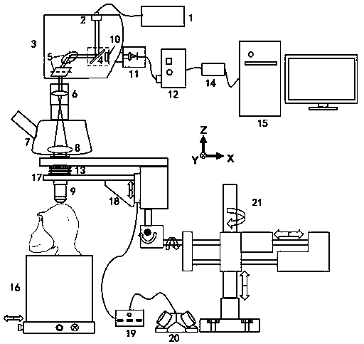
[0024] Example: such as figure 1 As shown, firstly, the laser light from the external laser 1 is extracted from the optical fiber, and then introduced into the confocal scanning module 3 through the collimator 2 . The confocal scanning module includes a dichroic mirror 4 (transmitting long wavelengths and reflecting short wavelengths) and two scanning galvanometers 5 whose rotation axes are perpendicular to each other. The dichroic mirror reflects the excitation light to the scanning galvanometer, which passes through Two reflections allow the excitation light to travel downwards. Afterwards, the excitation beam passes through the Scan lens 6 and the Tube lens 8 in the trinocular lens 7 in sequence, and then is expanded, and the expanded parallel light is focused into a point by the objective lens 9 to excite the probe. The generated fluorescence signal is collected by the objective lens, returns to the original path through the above optical path, and passes through the dich...


PUM


Abstract

The invention discloses a near-infrared two-zone confocal microscopic imaging system based on a multi-dimensional adjustment frame. According to the invention, a confocal microscope module is installed on a multi-dimensional adjustment frame, and an objective lens can be conveniently moved directly above the imaging plane through multi-dimensional translation and rotation. Near infrared laser is introduced from the port on a scanning unit and realizes scanning in the X-Y plane through a scanning galvanometer. Outgoing fluorescence is filtered out of focus signal in a pinhole module and finallydetected by a photomultiplier tube with near infrared two-zone response. The objective lens is installed on an electric focusing module, which can realize accurate focusing and tomography. Accordingto the invention, the multidimensional adjustment frame can be adopted to enable a confocal microscope to conveniently image large animals, and the performance is improved by optimizing the imaging band. The system of the invention has the advantages of excellent performance and simple use, and is suitable for promotion.

Description

technical field [0001] The invention belongs to the field of microscopic imaging of applied optics, and relates to a near-infrared two-zone confocal microscopic imaging system based on a multi-dimensional adjustment frame. Background technique [0002] Laser confocal scanning microscope is a high-precision microscopic imaging device developed on the basis of traditional fluorescence microscope. It adopts the mode of point excitation and point detection, and has high spatial resolution and signal-to-noise ratio. It plays an important role in the field of optical biological imaging. play an important role. In the structure of the confocal scanning microscope, the laser is used as the excitation light source and is focused on a point on the imaging plane by the objective lens, and the scanning on the X-Y plane is realized through a scanning module (such as a scanning galvanometer). There is a pinhole structure at the position where the front end of the receiving surface of the...

Claims


Application Information

Patent Timeline
Patent Type & Authority Applications(China)
IPC IPC(8): A61B5/02
CPCA61B5/0071A61B5/02007A61B2503/40
Inventor 钱骏蔡朝冲
Owner ZHEJIANG UNIV
Why Eureka
Try Eureka