Method and system for removing metal artifacts of oral cavity cone beam CT (Computed Tomography) image

A technology of metal artifacts and CT images, applied in the field of medical imaging, can solve the problems of discontinuous boundary information of metal areas, difficulty in application, and weaken secondary artifacts, so as to avoid boundary discontinuity and distortion of surrounding tissue information, and ensure accuracy Sex, the effect of suppressing secondary artifacts

Pending Publication Date: 2021-12-10
FUSSEN TECH CO LTD
View PDF0 Cites 0 Cited by
  • Summary
  • Abstract
  • Description
  • Claims
  • Application Information

AI Technical Summary

Problems solved by technology

However, the iterative reconstruction method mainly establishes a minimization model based on known constraints for iterative optimization, but the calculation time is long and it is difficult to apply in practice
The projection correction method mainly performs interpolation correction on the metal area of ​​the projection data, which is easy to implement and has a fast calculation speed. However, due to missing information, the boundary information of the metal area will be discontinuous and the surrounding tissue information will be distorted, and it is easy to introduce serious secondary artifacts; some The projection correction method corrects the projection of the metal area by calculating the prior image, which can reduce the secondary artifacts to a certain extent, but the effect still needs to be further improved to better meet the clinical diagnosis requirements

Method used


Image

  • Method and system for removing metal artifacts of oral cavity cone beam CT (Computed Tomography) image
  • Method and system for removing metal artifacts of oral cavity cone beam CT (Computed Tomography) image
  • Method and system for removing metal artifacts of oral cavity cone beam CT (Computed Tomography) image

Examples


Embodiment Construction

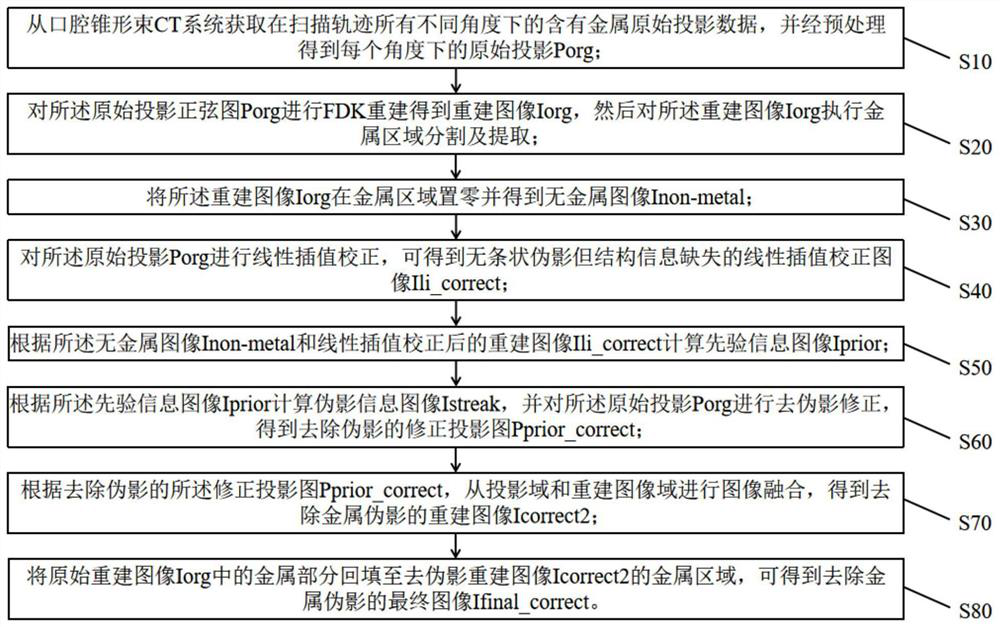
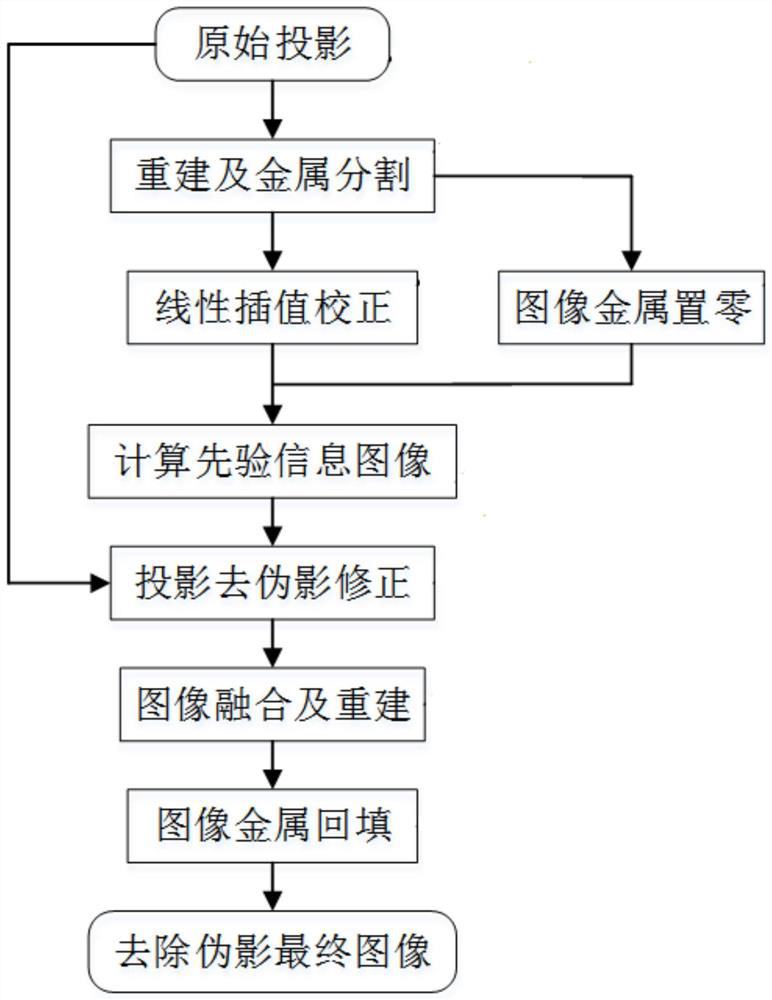
[0054] In order to make the object, technical solution and advantages of the present invention clearer, the present invention will be further described in detail below in conjunction with the accompanying drawings and embodiments. It should be understood that the specific embodiments described here are only used to explain the present invention, not to limit the present invention.

[0055] In the description of the present invention, it should be noted that unless otherwise specified and limited, the terms "installation", "connection" and "connection" should be understood in a broad sense, for example, it can be a fixed connection or a detachable connection. Connected, or integrally connected; it can be mechanically or electrically connected; it can be directly connected, or indirectly connected through an intermediary, or it can be the internal communication of two components, which can be wireless or wired connect. Those of ordinary skill in the art can understand the speci...


PUM


Abstract

The invention relates to the technical field of medical imaging, and discloses a method and system for removing metal artifacts of an oral cavity cone beam CT (Computed Tomography) imag and the method comprises the steps: obtaining an original projection Porg, a reconstruction image Iorg, a metal-free image Inon-metal and a linear interpolation correction image Ilicorrect, and carrying out the calculation to obtain a prior information image Iprior; calculating an artifact information image Istream according to the prior information image Iprior, and carrying out artifact removal correction on the original projection Porg, so that a corrected projection image Pprior_correct without artifacts is obtained; according to the corrected projection image Ppriorcorrect from which the artifacts are removed, carrying out image fusion from a projection domain and a reconstructed image domain to acquire a reconstructed image Icorrect2 from which the artifacts are removed; and backfilling the metal part in the original reconstructed image Iorg to the metal area of the artifact-removed reconstructed image Icorrect2, so that a final image Ifinalcorrect of which the metal artifacts are removed can be obtained. Metal artifacts can be removed in the oral cavity cone beam CT reconstruction, meanwhile, secondary artifacts are restrained, metal area boundary discontinuity and surrounding tissue information distortion are avoided, and the accuracy of oral cavity examination and diagnosis of doctors is guaranteed.

Description

technical field [0001] The invention relates to the technical field of medical imaging, in particular to a method and a system for removing metal artifacts of oral cavity cone beam CT images. Background technique [0002] CT tomography is one of the most routine medical examination methods in the development of modern medical imaging. During oral CBCT scanning, there are often high-density attenuating materials such as metal crowns, dentures and implants in the patient’s oral cavity, resulting in the appearance of reconstructed images. Severe metal artifacts. These high-density attenuating substances such as metals will harden the X-ray beam passing through them, the number of photons reaching the detector is insufficient, and the projection data at the junction of the metal and the tissue changes drastically and discontinuously. The problematic projections are filtered and back-projected There will be dark band artifacts, strip artifacts and pinstripe noise interference, t...

Claims


Application Information

Patent Timeline
Patent Type & Authority Applications(China)
IPC IPC(8): G06T11/00G06T7/136G06T7/11G06T5/30G06T5/00G06T3/40
CPCG06T11/008G06T11/005G06T7/11G06T5/30G06T7/136G06T5/002G06T3/4007G06T2207/10081
Inventor 李华勇黄俊杰李训青
Owner FUSSEN TECH CO LTD
Why Eureka
Try Eureka