Synthetically focused ultrasonic diagnostic imaging system for tissue and flow imaging

A focused ultrasound, ultrasound image technology, applied in ultrasound/sonic/infrasonic Permian technology, ultrasound/sonic/infrasound image/data processing, blood flow measurement devices, etc. Frame rate halving, etc.

Inactive Publication Date: 2005-08-03
KONINKLIJKE PHILIPS ELECTRONICS NV
View PDF4 Cites 17 Cited by
  • Summary
  • Abstract
  • Description
  • Claims
  • Application Information

AI Technical Summary

Problems solved by technology

However, each group of ultrasound beams focused differently increases the time required to obtain image information, which slows down the frame display rate
For example, advancing from one focal zone to two will roughly halve the frame rate

Method used


Image

  • Synthetically focused ultrasonic diagnostic imaging system for tissue and flow imaging
  • Synthetically focused ultrasonic diagnostic imaging system for tissue and flow imaging
  • Synthetically focused ultrasonic diagnostic imaging system for tissue and flow imaging

Examples


Embodiment Construction

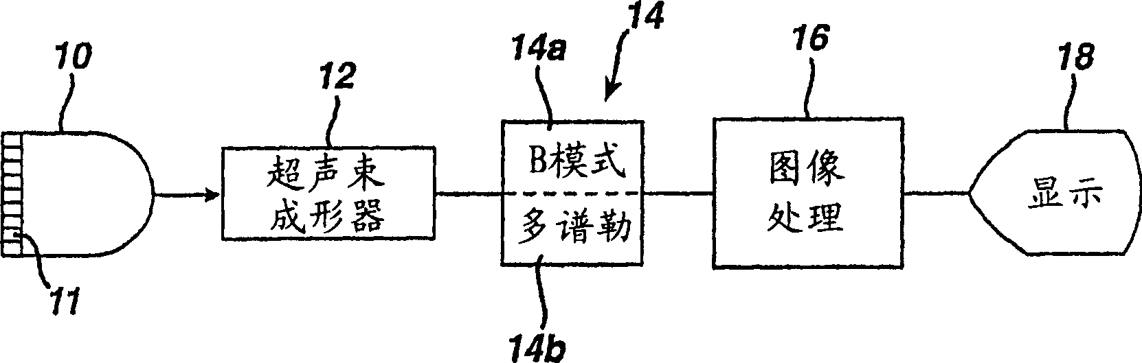
[0022] Current diagnostic ultrasound systems can be described as having 3 distinct modes of operation: B-mode echo (i.e., gray scale), spectral Doppler (i.e., single-gated Doppler, with high spectral resolution), and color Doppler (i.e., multi-gated Doppler, in which a single scalar quantity such as mean Doppler frequency or total Doppler power is evaluated in multiple sample volumes, but essentially no spectral resolution Rate). This division is reflected both in the architecture of the system and in the functionality of the system. In general, dedicated hardware is required to implement each mode, and only one mode can be operated at a time. An example of such a standard ultrasound system architecture is shown in FIG. 1 . The probe 10 has an array of transducers 11 which under the control of an ultrasound beam shaper 12 direct ultrasound beams along selected lines through the image field. Echoes are formed within the line of coherent echo signals along said line by an ult...


PUM


Abstract

A synthetic focus ultrasound system is described which is operated in a hybrid fashion. System operation alternates between synthetic focus acquisition and conventional focused beam acquisition. This makes possible, for example, the acquisition and display of harmonic images. Speckle artifacts in the synthetic focused ultrasound images may be reduced by combining signals from different sub-apertures which view the image field from different look directions. In a described embodiment, sets of motion maps are be produced for different sub-apertures of the array transducer, then compounded to reduce speckle. One or more identified regions of interest within a synthetic focused ultrasound image may be processed differently from other regions of the image to highlight or better define particular motional characteristics within the regions of interest, such as turbulent flow or different velocities of flow or motion.

Description

technical field [0001] The present invention relates to ultrasonic diagnostic imaging systems, and more particularly to synthetically focused ultrasonic diagnostic imaging systems that image tissue, blood flow, and blood flow patterns. Background technique [0002] Conventional ultrasound systems today produce images by scanning an area of ​​the body with an ultrasound beam that is formed electrically by an array of transducers. The timing of the signals applied to the array elements determines the direction in which the ultrasound beam is sent and the depth of focus of the ultrasound beam. Multiple adjacent ultrasound beams are sent to spatially adequately sample the target area. Echoes in the direction of the ultrasound beam are processed in known manner to form images of a plane or volume scanned by the ultrasound beam and to characterize tissue or blood flow in said plane or volume. [0003] When transmitting an ultrasound beam, the beam profile determined by the timin...

Claims


Application Information

Patent Timeline
Patent Type & Authority Applications(China)
IPC IPC(8): A61B8/06A61B8/08G01S7/52G01S15/89
CPCG01S15/8979G01S15/8927G01S15/8918G01S7/52028G01S15/8977G01S15/8925
Inventor B·S·罗宾森C·R·库利
Owner KONINKLIJKE PHILIPS ELECTRONICS NV
Why Eureka
Try Eureka