
Protein sequencing methods and reagents

a technology of protein and reagent, applied in the field of protein sequencing, can solve the problems of inapplicability to heterogeneous samples, low sensitivity of technology, and serious constraints of existing techniques, and achieve the effect of facilitating the chemical cleavage of the n-terminal amino acid

Active Publication Date: 2022-03-08
THE GOVERNING COUNCIL OF THE UNIV OF TORONTO
View PDF7 Cites 1 Cited by
  • Summary
  • Abstract
  • Description
  • Claims
  • Application Information

AI Technical Summary

Benefits of technology

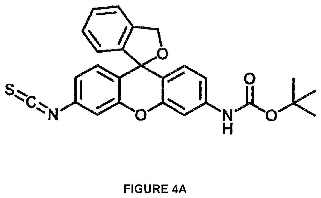
[0028]In one embodiment, the probe comprises a synthetic fluorescent dye. In one embodiment, the probe comprises a xanthene derivative, such as a dye based on fluorescein, eosin, or rhodamine. For example, in one embodiment, the probe comprises hydroxymethyl rhodamine green (HMRG). In one embodiment, the probe is suitable for optical detection using super-resolution microscopy. In one embodiment, the probe is a spontaneously blinking dye or a photoswitchable dye. In one embodiment, the probe is facilitates the chemical cleavage of the N-terminal amino acid from the polypeptide. For example, in some embodiment the probe comprises a reactive isothiocyanate (ITC) group.

Problems solved by technology

Yet while methods for protein identification, quantification and imaging are needed throughout biomedicine, existing techniques have serious constraints.
While automatable, the technology has low sensitivity and is not applicable to heterogeneous samples (i.e. mixtures of different polypeptides), limiting utility.
Yet it also suffers from limited dynamic range, biased detection, and incremental performance gains.
Moreover, it is based on ensemble measurements (bulk analysis), resulting in the loss of valuable contextual information (e.g. protein subcellular localization etc.).
Crucially, however, these methods depend on the availability of fluorescent antibodies, or other similarly labeled affinity capture reagents such as aptamers, which are physically large relative to most cellular polypeptides, diminishing resolution, and / or which may bind cellular proteins in addition to the target of interest, producing artifacts, and typically fewer than 3-4 different cellular proteins can be imaged together at one time.

Method used


Image

  • Protein sequencing methods and reagents
  • Protein sequencing methods and reagents
  • Protein sequencing methods and reagents

Examples


example 1

lecule Spectroscopy-Based Amino Acid Residue Identification

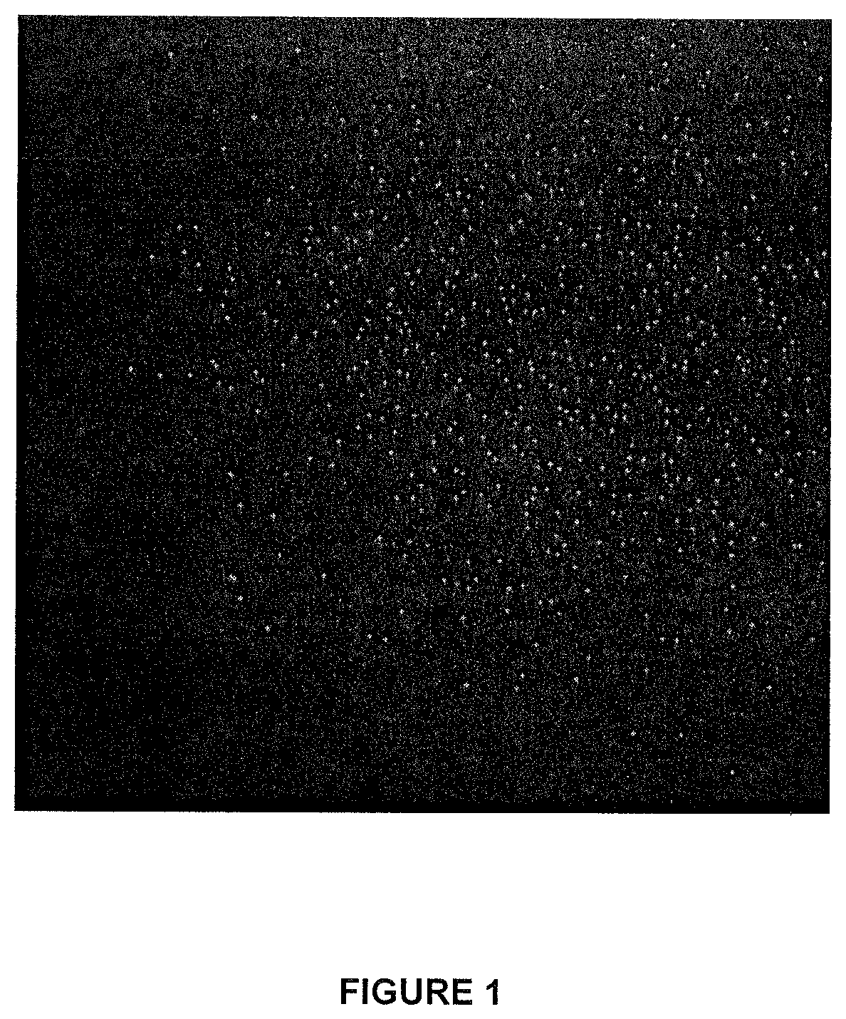
[0074]The inventor has adapted super-resolution fluorescence microscopy to an Edman-like sequencing process, allowing for the simultaneous, massively parallel identification and counting of large numbers of individual protein molecules. The protein molecules may be in vitro or in situ such as in cells and / or tissues. N-terminal amino acid residues of affixed polypeptides are reacted with a fluorescent probe that confers distinct spectral properties upon coupling to different amino acids. The resulting characteristic emission profile generated by the distinct N-terminal derivative formed on individual protein molecules is monitored by super-resolution spectroscopy to determine the identity of the corresponding cognate amino acid residue, which is then cleaved off, such as through Edman-like chemistry. Through multiple, iterative cyclic rounds of coupling the remaining polypeptide portions to fresh dye, re-imaging the populati...

example 2

ation and Characterization of Fluorescent Probes

[0082]Using fluorescent probes some, or all, of the 20+ naturally occurring amino acids may be distinguished by the detecting the fluorescent emission of the individual probes conjugated to different amino acids. Various dye molecules, such HMRG and other xanthene-derivatives, are investigated for their suitability as probes for single molecule spectroscopy-based residue identification.

[0083]Experiments are conducted in order to identify fluorescent probes suitable for distinguishing different amino acids and to define optimal imaging conditions (eg. buffer pH / polarity) that maximize discrimination, as well as the influence, if any, of adjacent amino acids (eg. penultimate residue). Large numbers of individual probe molecules are examined after immobilization to a surface (eg. coverslip, flowcell, or microbead), or natively in / on metazoan cells, microbes or virus, in order to derive a precise, imaging-based N-terminal readout for one, ...

example 3

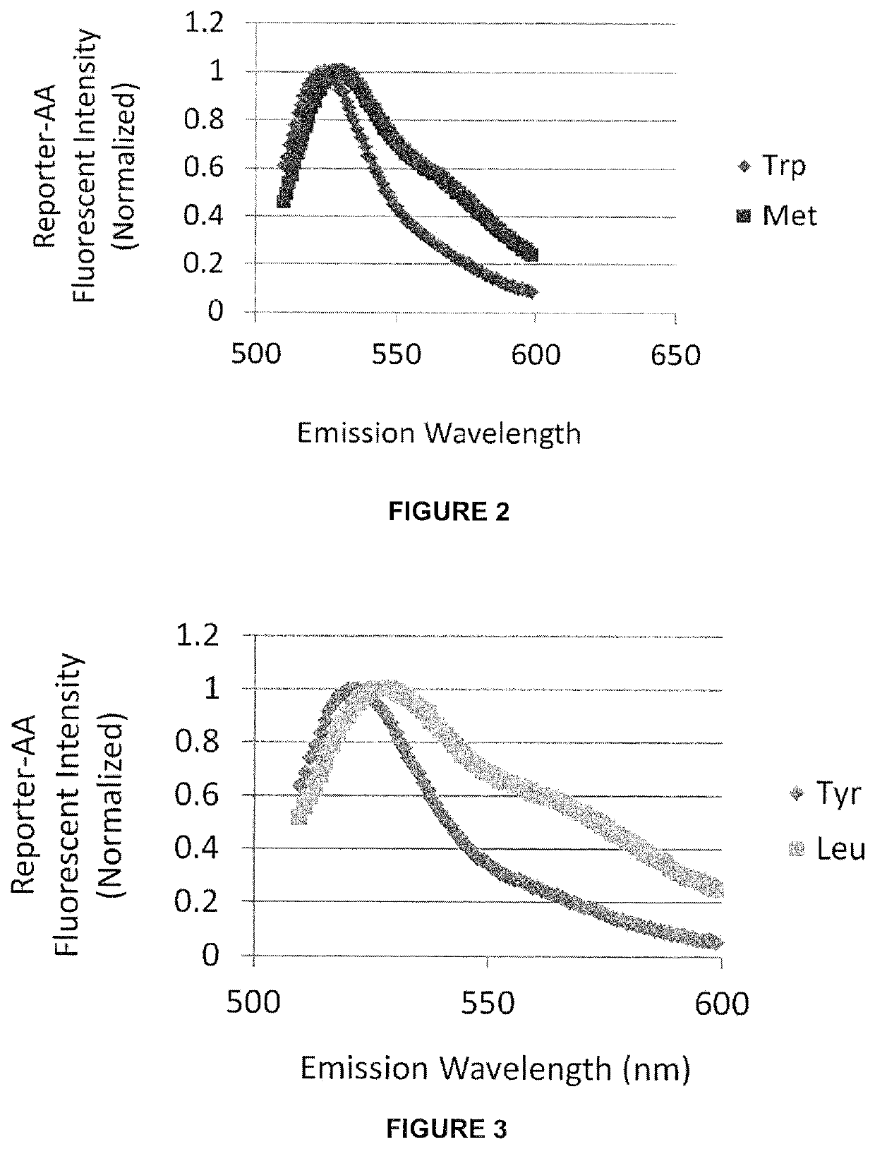
ITC Coupling and Identifying Different N-Terminal Amino Acid by Fluorescence Profiling

Materials and Methods

[0086]Hydroxymethyl rhodamine green (HMRG) tert-butyloxycarbonyl (BOC) isothiocyanate (ITC) (HMRG-BOC-ITC) as shown in FIG. 4A was synthesized as shown in FIG. 4B and dissolved in DMSO to a final concentration 34 nmol / μl.

[0087]Peptide beads: Peptide beads were synthesized by Kinexus (Vancouver B.C). Tentagel resin (Tenta-Gel® M NH2, 10 μm beads, RAPP Polymer) was used as the support for solid peptide synthesis. The peptides were synthesized to have the amino acid sequence X-AGWYMRLG (SEQ ID NO: 1), where X represents any one of 20 different amino acids at N-termini of the peptide. Approximately half (˜50%) of each peptide / resin contained a cleavable HMBA linker inserted between peptide sequence and the bead which enabled cleavage of the peptide molecules from the beads upon treatment with base.

[0088]Prior to coupling, 100 μl of dimethylformamide (DMF) was added to each tube con...


PUM

PropertyMeasurementUnit
pHaaaaaaaaaa
surface areaaaaaaaaaaa
slit widthaaaaaaaaaa

Abstract

Described are optical methods and reagents for sequencing polypeptides. A probe that exhibits different spectral properties when conjugated to different N-terminal amino acids is conjugated to the N-terminal amino acid of a polypeptide. Sequentially detecting one or more spectral properties of the probe conjugated to the N-terminal amino acid and cleaving the N-terminal amino acid produces sequence information of the polypeptide. The use of super-resolution microscopy allows for the massively parallel sequencing of individual polypeptide molecules in situ such as within a cell. Also described are probes comprising hydroxymethyl rhodamine green, an isothiocyanate group and a protecting group.

Description

RELATED APPLICATIONS[0001]This application is a national phase entry of PCT / CA2016 / 051204 filed Oct. 17, 2016 (which designates the U.S.) which claims the benefit of priority of U.S. Provisional Patent Application No. 62 / 242,619 filed Oct. 16, 2015, the contents of which are hereby incorporated by reference.INCORPORATION OF SEQUENCE LISTING[0002]A computer readable form of the Sequence Listing “13795-P49671US01 SequenceListing.txt” (4,096 bytes), submitted via EFS-WEB and created on Apr. 13, 2018, is herein incorporated by reference.FIELD OF THE INVENTION[0003]This invention relates to the field of protein sequencing and more specifically to methods, assays and reagents for sequencing protein or polypeptide molecules as well as to fluorescent single molecule imaging methods, assays and reagents for sequencing individual polypeptide molecules.BACKGROUND OF THE INVENTION[0004]Proteins underlie virtually every biological process; perturbations in their expression, degradation, interact...

Claims


Application Information

Patent Timeline
Patent Type & Authority Patents(United States)
IPC IPC(8): G01N31/00G01N33/53G01N33/68C07D493/10C40B20/04
CPCG01N33/6824C07D493/10C40B20/04Y02P20/55
Inventor EMILI, ANDREW
Owner THE GOVERNING COUNCIL OF THE UNIV OF TORONTO
Learn More