Methods and devices for endosonography-guided fundopexy

a technology of endoscopy and fundopexy, which is applied in the field of tissue securement system, device and method for endoscopy or endosonographyguided transluminal intervention, can solve the problems of high incidence of postoperative complications, extended recovery time, and limited endoscopic treatment, and achieve the effect of reducing gastroesophageal reflux

Active Publication Date: 2006-12-14
ADVENT MEDICAL
View PDF104 Cites 46 Cited by
  • Summary
  • Abstract
  • Description
  • Claims
  • Application Information

AI Technical Summary

Benefits of technology

[0008] Accordingly, an objective of this invention is to provide a method to reduce gastroesophageal reflux by endosonography-guided intervention. Specifically, endosonography is used to insert a ligation element through the esophageal wall, through the diaphragmatic crus and into the fundus of the stomach. This ligation element placed from the esophagus and around the angle of His may create a barrier to gastroesophageal reflux.
[0011] In another aspect of the invention, the system for fastening tissue also includes a ligating element that can be attached to the proximal end of the connecting element once the hollow needle is removed. The ligating element may utilize a dilating element positioned at its distal end that is sized to dilate a tissue tunnel so that the ligating element may be drawn more easily through the tissue structures.
[0012] In still another aspect of the invention, the system may also utilize an endoscopically guided grasper to grasp the distal end of a connecting element and pull the ligating element across the esophageal wall and into the stomach. The system may also utilize a securement element that is configured to engage the proximal and distal ends of the ligating element together. This may prevent the ligating element loop from loosening.

Problems solved by technology

However this procedure can have a high incidence of postoperative complications and extended recovery times. Therefore endoscopic methods for enhancing the lower esophageal sphincter have been developed as an alternative to surgery.
Endoscopic treatment is often limited because the operator can only visualize the mucosal lining of the gastrointestinal wall that is located directly in front of the endoscope.

Method used


Image

  • Methods and devices for endosonography-guided fundopexy
  • Methods and devices for endosonography-guided fundopexy
  • Methods and devices for endosonography-guided fundopexy

Examples


Embodiment Construction

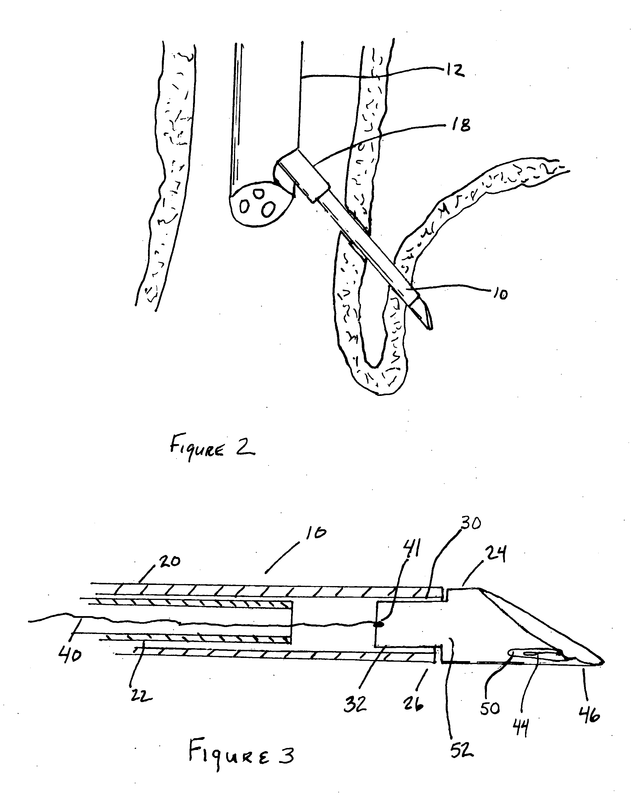
[0025] The system and method described herein may offer improvements over the techniques currently utilized to perform endoscopic procedures. This tissue securement system has several embodiments that are intended to work together to create a novel device and method for the treatment of GERD. However these embodiments also function independently and some of the embodiments of this system may be removed and the system may still achieve its desired function. Alternatively several of these embodiments may be useful as stand alone devices. The principle elements of this system are; an echoendoscope, a delivery needle, a stylet, a needle tip, a connecting element, a ligating element and securement elements.

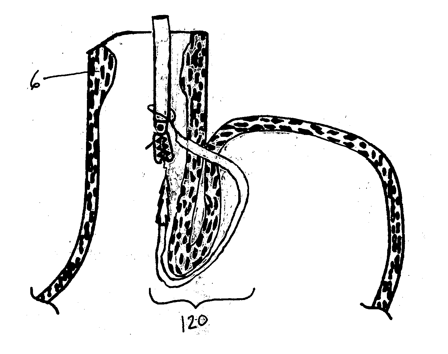
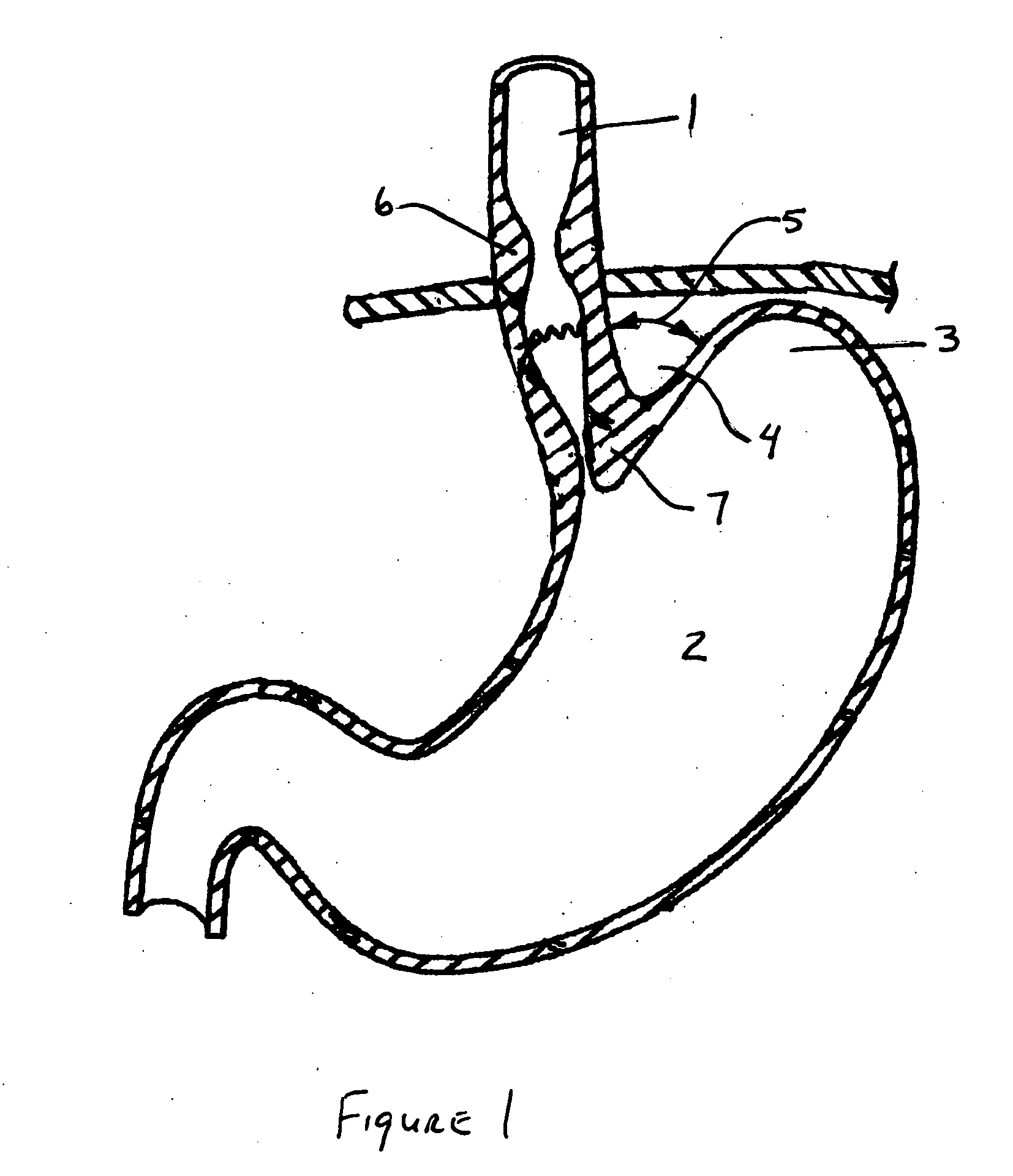
[0026]FIG. 1 is a cross sectional view of the esophageal-gastro-intestinal tract shown from the esophagus 1 to the stomach 2. The fundus 3 forms the superior portion of the stomach 2. The esophagus 1 enters the stomach 2 at a point below the fundus 3 forming the cardiac notch 4 and an...


PUM


Abstract

The present invention relates to a tissue securement system, device and method for endoscopy or endosonography-guided transluminal interventions whereby a ligation or anchor is placed and secured into soft tissue. An objective of this invention is to provide a method to reduce gastroesophageal reflux by endosonography-guided intervention. Specifically, endosonography is used to insert a ligation element through the esophageal wall, through the diaphragmatic crus and into the fundus of the stomach. This ligation element placed from the esophagus and around the angle of His that may create a barrier to gastroesophageal reflux.

Description

CLAIM OF PRIORITY [0001] This application claims priority under 35 U.S.C. § 119(e) to U.S. Provisional Application No. 60 / 688,837, filed Jun. 9, 2005, the entire contents of which are hereby expressly incorporated by reference.BACKGROUND [0002] 1. Field of the Invention [0003] The present invention relates to a tissue securement system, device and method for endoscopy or endosonography-guided transluminal interventions whereby a ligation or anchor is placed and secured into soft tissue. [0004] 2. Background of the Invention [0005] Gastroesophageal reflux disease (GERD) is a chronic condition caused by the failure of the anti-reflux barrier located at the gastroesophageal junction to keep the contents of the stomach from refluxing back into the esophagus. [0006] Surgical fundoplication is the gold standard for anatomic correction of the cardia in patients with GERD. However this procedure can have a high incidence of postoperative complications and extended recovery times. Therefore ...

Claims


Application Information

Patent Timeline
Patent Type & Authority Applications(United States)
IPC IPC(8): A61B17/12
CPCA61B17/12013A61B2017/003A61B17/0401A61B2017/0409A61B2017/00827
Inventor BINMOELLER, KENNETH F.
Owner ADVENT MEDICAL
Why Eureka
Try Eureka