Methods and devices for facilitating visualization in a surgical environment

Active Publication Date: 2008-04-03
ACCLARENT INC
View PDF61 Cites 137 Cited by
  • Summary
  • Abstract
  • Description
  • Claims
  • Application Information

AI Technical Summary

Benefits of technology

[0037]In at least one embodiment, the distal end portion of the illuminating device is advanced further distally of a distal end of the sinus guide

Problems solved by technology

If the mucosal tissue of one of these passageways becomes inflamed for any reason, the cavities which drain through that passageway can become blocked.
This interference with drainage of mucous (e.g., occlusion of a sinus ostium) can result in mucosal congestion within the paranasal sinuses.
Chronic mucosal congestion of the sinuses can cause damage to the epithelium that lines the sinus with subsequent decreased oxygen tension and microbial growth (e.g., a sinus infection).
A large number of patients do not respond to drug therapy.
For example, FESS can cause significant post-operative pain.
Also, some FESS procedures are associated with significant postoperative bleeding and, as a result, nasal packing is frequently pl

Method used


Image

  • Methods and devices for facilitating visualization in a surgical environment
  • Methods and devices for facilitating visualization in a surgical environment
  • Methods and devices for facilitating visualization in a surgical environment

Examples


Example

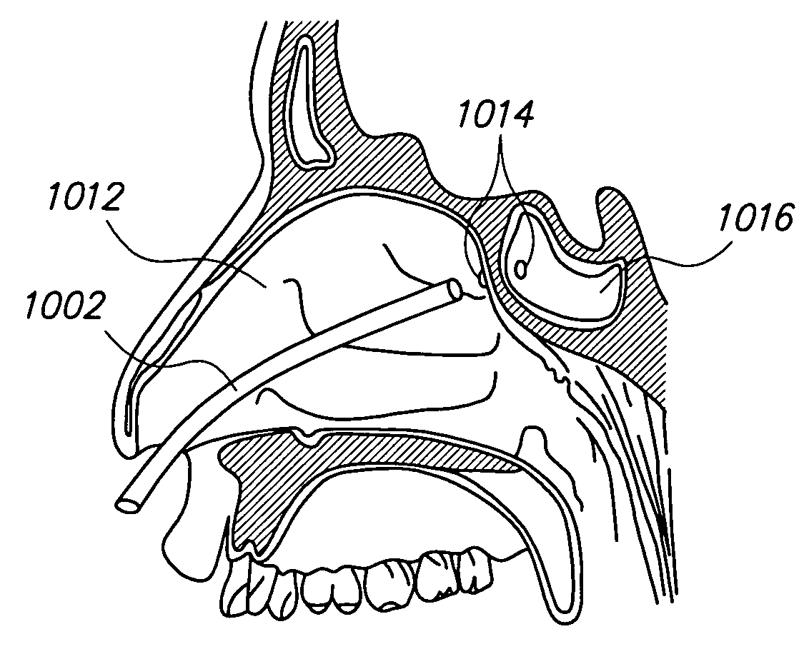
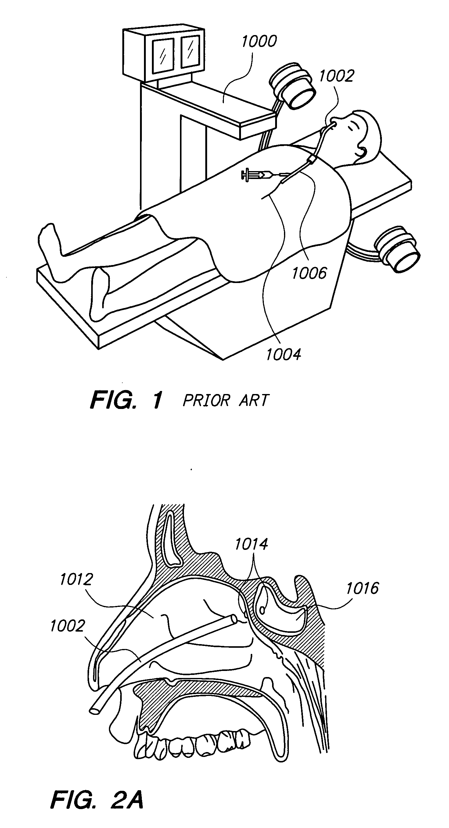
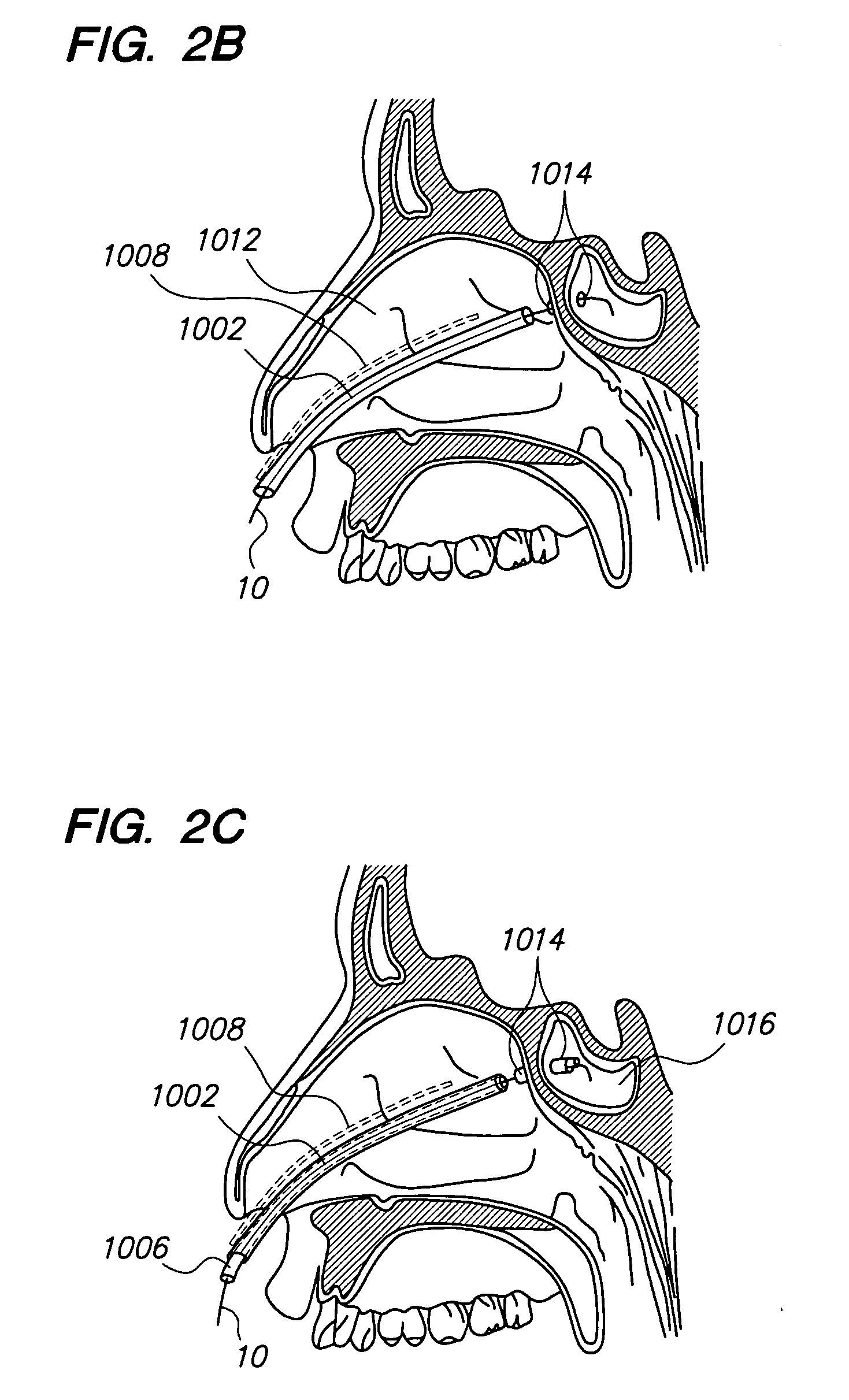
[0160]Before the present devices and methods are described, it is to be understood that this invention is not limited to particular embodiments described, as such may, of course, vary. It is also to be understood that the terminology used herein is for the purpose of describing particular embodiments only, and is not intended to be limiting, since the scope of the present invention will be limited only by the appended claims.

[0161]Where a range of values is provided, it is understood that each intervening value, to the tenth of the unit of the lower limit unless the context clearly dictates otherwise, between the upper and lower limits of that range is also specifically disclosed. Each smaller range between any stated value or intervening value in a stated range and any other stated or intervening value in that stated range is encompassed within the invention. The upper and lower limits of these smaller ranges may independently be included or excluded in the range, and each range wh...


PUM


Abstract

Devices and methods for visually confirming the positioning of a distal end portion of an illuminating device placed within a patient include inserting a distal end portion of an illuminating device internally into a patient, emitting light from the distal end portion of the illuminating device, observing transillumination resulting from the light emitted from the distal end portion of the illuminating device that occurs on an external surface of the patient, and correlating the location of the observed transillumination on the external surface of the patient with an internal location of the patient that underlies the location of observed transillumination, to confirm positioning of the distal end portion of the illuminating device.

Description

FIELD OF THE INVENTION[0001]The present invention relates generally to medical devices, systems and methods and more particularly to methods and devices for performing minimally invasive procedures that reduce the need to provide fluoroscopic or other radiographic visualization.BACKGROUND OF THE INVENTION[0002]The skull contains a series of cavities known as paranasal sinuses that are connected by passageways. The paranasal sinuses include frontal sinuses, ethmoid sinuses, sphenoid sinuses and maxillary sinuses. The paranasal sinuses are lined with mucous-producing mucosal tissue and ultimately open into the nasal cavity. Normally, mucous produced by the mucosal tissue slowly drains out of each sinus through an opening known as an ostium. If the mucosal tissue of one of these passageways becomes inflamed for any reason, the cavities which drain through that passageway can become blocked. This blockage can be periodic (resulting in episodes of pain) or chronic. This interference with...

Claims


Application Information

Patent Timeline
IPC IPC(8): A61M29/00
CPCA61B1/00126G02B6/4292A61B5/0084A61B5/061A61B5/064A61B5/6851A61B2562/228A61M25/0105A61M25/09A61M2025/09083A61M2025/09125A61M2025/09175G02B6/0006G02B6/04A61B2018/2255A61B1/07G02B6/262G02B6/241G02B6/1228G02B6/125A61B1/233A61B2018/2222A61B2018/2244A61B34/20A61B2034/2055A61B2090/306A61B1/063A61B1/0684A61B5/062A61B17/24A61B2017/246A61B2217/005A61M29/02A61M2025/0166A61M2025/09008A61M2025/09166A61M2029/025G02B6/0008G02B23/2469
Inventor GOLDFARB, ERICMORRISS, JOHNCHANG, JOHN Y.FACTEAU, WILLIAM M.
Owner ACCLARENT INC
Why Eureka
Try Eureka