Radiotherapy support apparatus

a support apparatus and radiotherapy technology, applied in the direction of electrical apparatus, radiation therapy, therapy, etc., can solve the problems of not being able to completely assure the planned irradiation of the patient, difficult to place the patient in a position according to the irradiation plan, and no means for confirming whether irradiation is actually performed, etc., to achieve accurate and safe operation

Active Publication Date: 2009-07-09
TOSHIBA MEDICAL SYST CORP
View PDF8 Cites 76 Cited by
  • Summary
  • Abstract
  • Description
  • Claims
  • Application Information

AI Technical Summary

Benefits of technology

[0008]In view of the circumstances, an object of the present invention is to provide an apparatus for supporting radiotherapy treatment to enable treatment to be performed accurately and safely by providing the operator with information for preventing insufficient irradiation of a region to be treated and excessive irradiation of a normal tissue in radiotherapy treatment.

Problems solved by technology

However, at present, there is no means for confirming whether irradiation is actually performed to the patient with the planned position and dose or not.
However, it is difficult to place a patient in a position according to the irradiation plan, on a bed different from a phantom which is easily portable and whose position can be freely adjusted.
The confirmation prior to radiation does not completely assure the planned irradiation of the patient.
Since forward scattering is dominant in the scattering in a subject of a therapeutic beam (a few MeV) having high energy, when a detector is disposed in the incident X-ray direction, it is difficult to distinguish scattering radiation and penetrated radiation from each other.
Amid mounting social concern over medical errors, also in radiotherapy treatment, excessive irradiation of a patient has been reported and has become an issue.
Therefore, if the therapeutic X-ray beam is off from the irradiation target, normal tissue is considerably damaged.

Method used


Image

  • Radiotherapy support apparatus
  • Radiotherapy support apparatus
  • Radiotherapy support apparatus

Examples


first example

[0118](First Example)

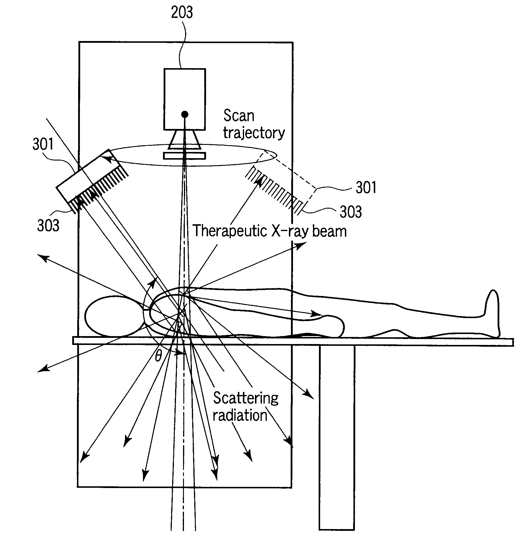
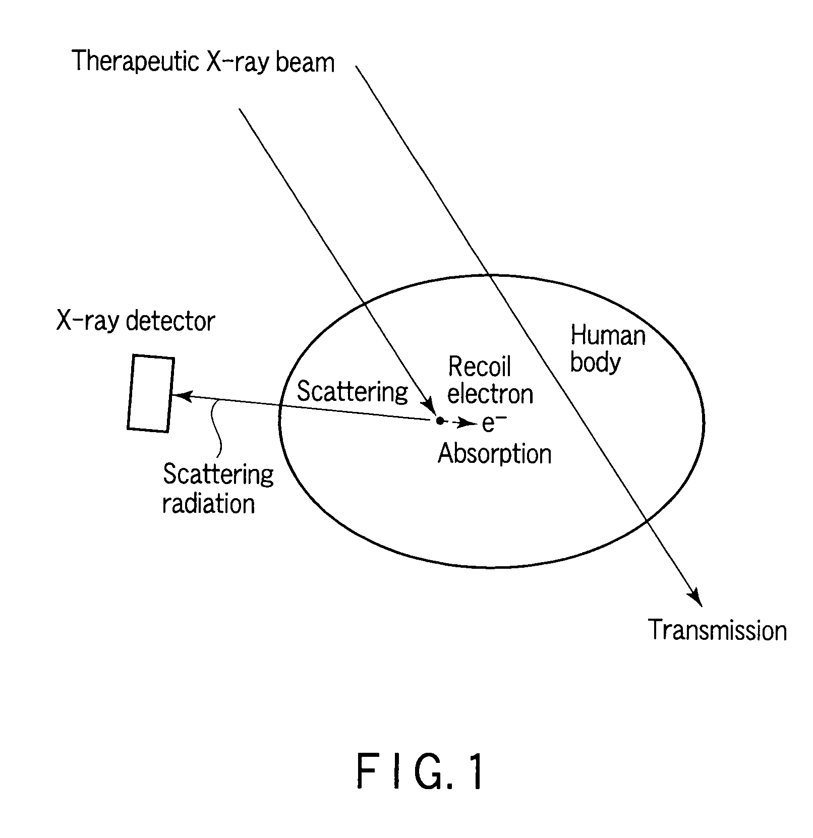
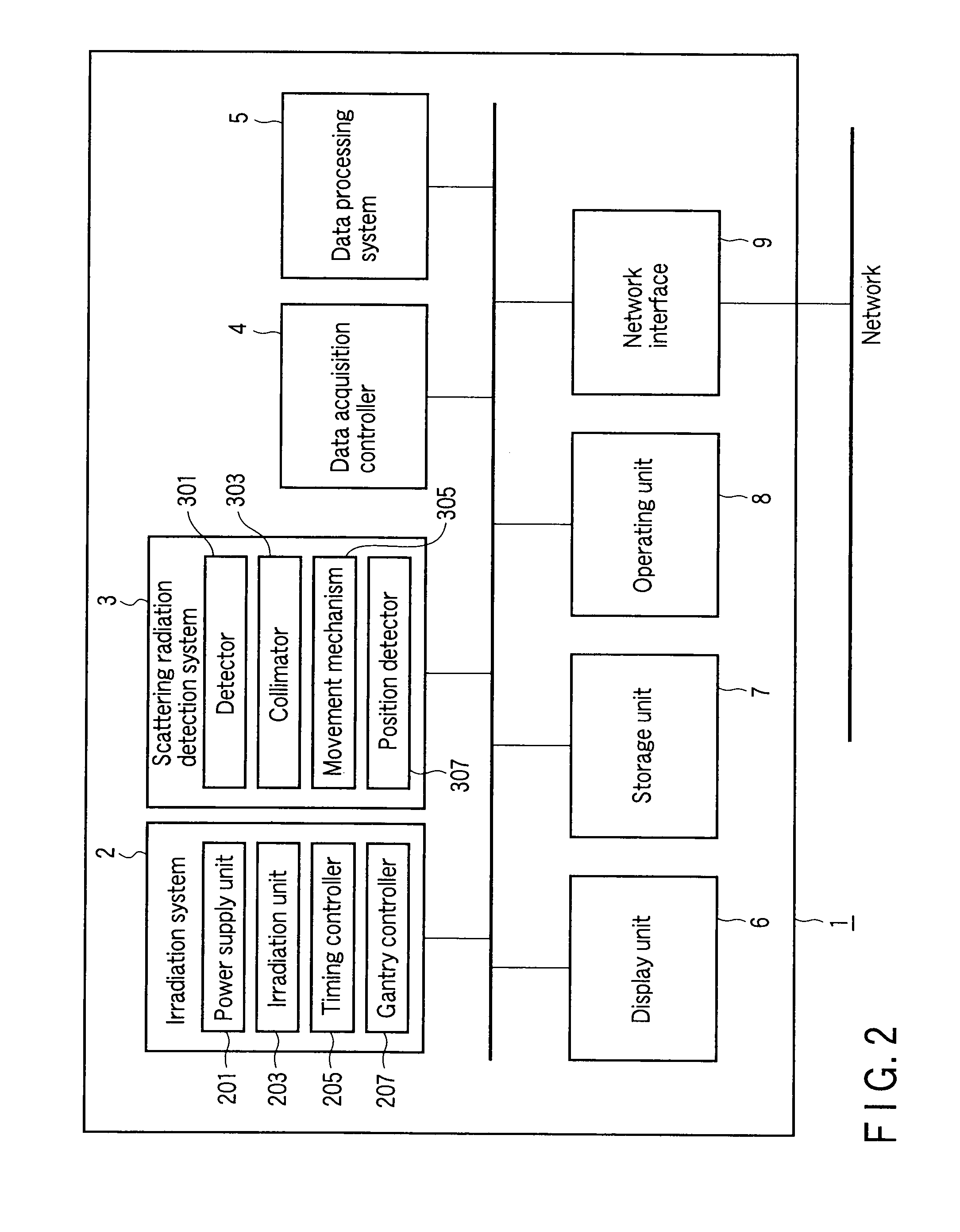
[0119]Next, a method of generating absorption dose image data using the radiotherapeutic system 1 of the first example will be described. In the radiotherapeutic system according to the first example, a detector having a collimator is mounted in a position at a specific angle with respect to a therapeutic X-ray beam and selectively detects only scattering radiation in the direction. Further, to three-dimensionally obtain a distribution of places where scattering occurs in the patient body, the detector is rotated during irradiation and scattering radiation is measured from all of directions (refer to, for example, FIG. 6). After that, a reconstruction process is performed, and a distribution of occurrence of scattering radiation in the subject is three-dimensionally imaged.

[0120]FIG. 5 is a flowchart showing the flow of processes in radiotherapy treatment including a process of generating absorption dose image data according to the example. The processes in the ...

second example

[0137](Second Example)

[0138]Next, a method of generating absorption dose image data using the radiotherapeutic system 1 of a second example will be described. In the radiotherapeutic system as the second example, a detector having a collimator is mounted in a position at a specific (scattering angle) angle with respect to a therapeutic X-ray beam and selectively detects only scattering radiation in the direction. By executing the detection while moving the therapeutic X-ray beam and the detection face and maintaining the angle formed between the axis of the therapeutic X-ray beam emitted from the irradiation unit and the detection face of the detector, a three-dimensional region in the subject is scanned. Scattering radiation volume data is reconstructed by using obtained three-dimensional scattering radiation data at the predetermined scattering angle, the scattering radiation volume data is converted to absorption dose volume data indicative of a three-dimensional distribution of ...

example 1

[0170][Display Example 1]

[0171]FIGS. 12A to 12C show an example of an absorption dose image displayed by the fusion model. Images in the upper stage of FIGS. 12A to 12C are cross sections perpendicular to the irradiation direction of the therapeutic radiation beam. Images in the lower stage are cross sections horizontal to the irradiation direction of the therapeutic radiation beam. FIG. 12A shows an example of display including a tumor (therapy target region) on which a radiation irradiated part and an accumulated absorption dose are superimposed. FIG. 12C displays a tissue (risk organ: risk region) existing near a therapeutic radiation beam passing region and relatively weak to irradiation. By displaying the irradiation history and the accumulated value, whether radiation does not exceed exposure limit or not can be confirmed on site. FIG. 12B shows an example of display where the images of FIGS. 12A and 12C are superimposed.

[0172]Although each of FIGS. 12A to 12C shows an example...


PUM


Abstract

A radiotherapy treatment support apparatus includes a storage unit which stores absorption dose volume data expressing a spatial distribution of absorption dose in a subject, a generation unit which generates fusion data associated with morphology volume data of the subject and the absorption dose volume data so as to be associated with a plurality of segments, and a display unit which displays an image which has the distribution of absorption dose superimposed on the two-dimensional morphology image of the subject using the fusion data.

Description

CROSS-REFERENCE TO RELATED APPLICATIONS[0001]This application is based upon and claims the benefit of priority from prior Japanese Patent Applications No. 2008-002370, filed Jan. 9, 2008; No. 2008-002371, filed Jan. 9, 2008; and No. 2008-002372, filed Jan. 9, 2008, the entire contents of all of which are incorporated herein by reference.BACKGROUND OF THE INVENTION[0002]1. Field of the Invention[0003]The present invention relates to an apparatus for supporting an operator at the time of radiotherapy treatment.[0004]2. Description of the Related Art[0005]In radiotherapy treatment typified by external X-ray radiotherapy treatment, before the treatment, an irradiation plan (the direction and the dose of radiation to an affected region) is made on a patient image. On the basis of the irradiation plan, irradiation of the patient is performed. However, at present, there is no means for confirming whether irradiation is actually performed to the patient with the planned position and dose or...

Claims


Application Information

Patent Timeline
Patent Type & Authority Applications(United States)
IPC IPC(8): H05G1/64
CPCA61N5/1048A61N2005/1054A61N2005/1052
Inventor SAKURAI, YASUOTAKADA, YOICHIKUMAKURA, MASAHIROOHYU, SHIGEHARUHARAGASHIRA, MOTOJINAMBU, KYOJIROSHIBATA, MARIKO
Owner TOSHIBA MEDICAL SYST CORP
Why Eureka
Try Eureka