Displaying Image Data From A Scanner Capsule

a scanner and image data technology, applied in the field of medical diagnostics, can solve the problems of increasing the incidence of cardiac problems in women, the current stretched and failed resources of physicians and health care professionals, and the limited number of current testing and monitoring systems

Inactive Publication Date: 2011-01-06
INNURVATION
View PDF112 Cites 121 Cited by
  • Summary
  • Abstract
  • Description
  • Claims
  • Application Information

AI Technical Summary

Benefits of technology

[0015]Images communicated outside of the patient represent tissues at exact locations determined based on signals transmitted. Such images can be communicated by radio wave, optically (such as, for example, using an optical fiber), by acoustic signals, etc. Signals representing images are received outside the patient's body and are processed by one or more computers running software capable of discarding duplicate information and stitching together, line scan by line scan a complete GI tract as if it were stretched out in a straight line. The processed, fully linear image is then displayed to a medical professional as if the GI tract had been stretched into a straight line, cut open, laid flat out on a bench for viewing—all without making any incisions in a live patient, as easy as swallowing a pill.

Problems solved by technology

There is a rising incidence of cardiac problems for women (the #1 cause of death for women).
Physician and health care resources are currently already stretched and will fail if the current technology, process and procedure are not altered to suit the needs of the baby boomer market of the near future.
Many current testing and monitoring systems are limited by old technology and processes that take days, if not weeks, for results.
These test methods, if not inconvenient and potentially embarrassing, are at least in most cases intrinsically painful or risky to patients.
Current camera pill usage by patients and physicians is limited for several reasons.
First and foremost, current technology is very large in comparison to most ingestible medicines and nutritional supplements.
The excessive size is in part a result of the selection of power-inefficient communication methods.
The large size mandates pre-screening of patients (an additional process, inconvenience, and cost).
The large size also leads to a reasonably high potential that the device can become lodged within the GI tract.
This may lead to a highly invasive surgical removal requirement, which carries all the risks associated with some surgeries.
Thus, current RF camera pills require at least two trips to the physician's office, as well as the wearing of a cumbersome belt reader with leads attached to the skin.
This diagnostic process is both inconvenient and uncomfortable.
It also carries a risk of surgical removal, due to the size of the current camera pills.
Current technology does not offer a recorded position within the body associated to the specific image taken.
Furthermore, the current camera pills are expensive devices, and are resorted to when other bowel disease diagnostic techniques, such as endoscopy and colonoscopy (each of which are extremely intrusive), present results that need further investigation.
Further, the electromagnetic signals used to transmit the images may harm the sensitive tissue they pass through in order to be detected outside the body.
Therefore, the current ingestible camera pill has significant deficiencies.
Due to the natural movement of the digestive tract, the resultant movies from the current camera pills depict spurts of forward and backward movements that are awkward and not reviewer friendly, leading to issues of reviewer focus and overall system effectiveness.

Method used


Image

  • Displaying Image Data From A Scanner Capsule
  • Displaying Image Data From A Scanner Capsule
  • Displaying Image Data From A Scanner Capsule

Examples


Embodiment Construction

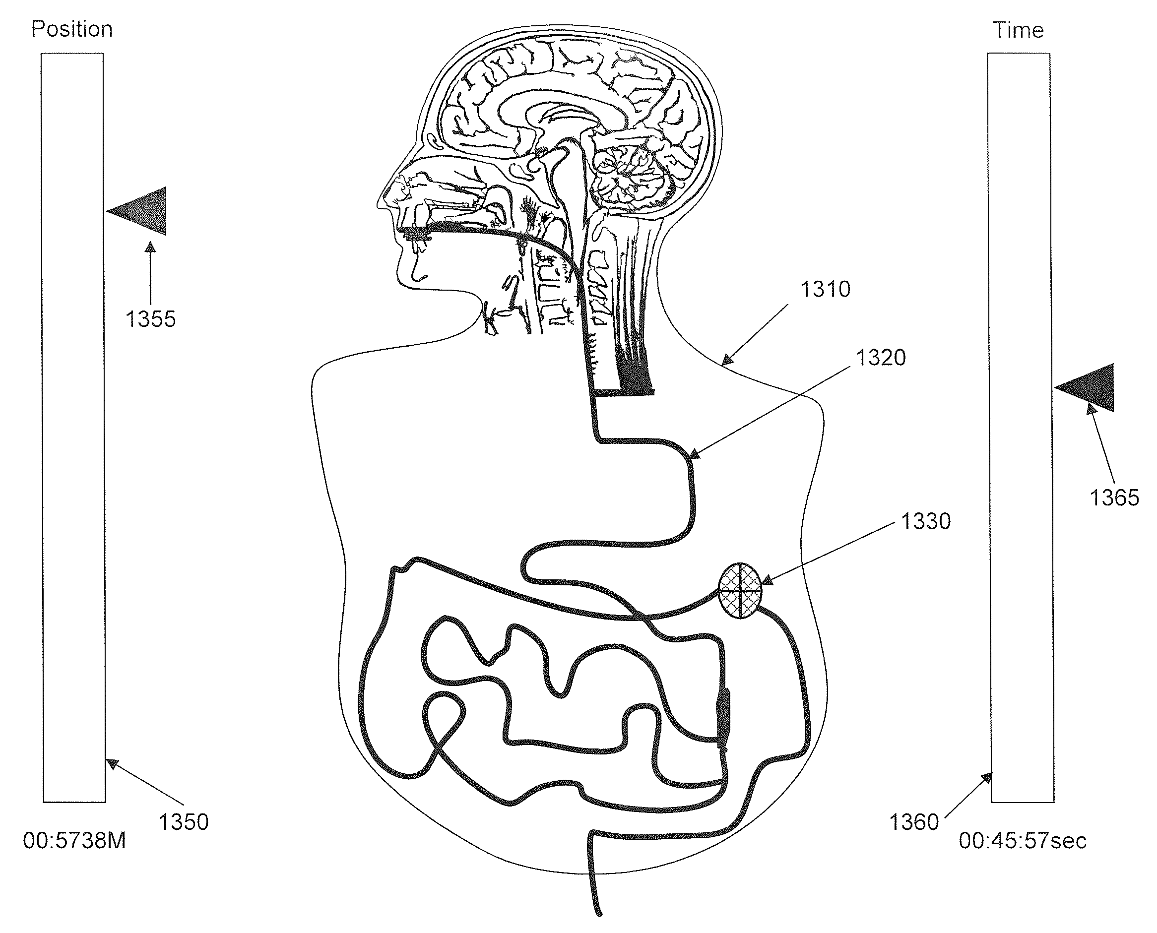
as depicted in FIG. 11.

[0038]FIG. 19 includes flow charts of subroutines of the image processing software for updating a tract aspect and updating time and distance, respectively, of the displays depicted in FIG. 11.

[0039]FIG. 20 is a flow chart of a subroutine of image processing software for updating a zoom aspect.

[0040]FIG. 21 is a flow chart of subroutines of image processing software for updating a tube aspect.

[0041]FIG. 22 is a flow chart of an overall scanned image collection, processing, and reporting system.

[0042]FIG. 23 is a detailed flow chart of a scanned image creation process.

[0043]FIG. 24 is an exemplary depiction of scanned data corresponding to FIG. 23 process.

[0044]Features and advantages of the present invention will become more apparent from the detailed description set forth below when taken in conjunction with the drawings, in which like reference characters identify corresponding elements throughout. In the drawings, like reference numbers generally indicate i...


PUM


Abstract

An ingestible image scanning pill captures high resolution images of the GI tract as it passes through. Images communicated externally have exact location determination. Image processing software discards duplicate information and stitches images together, line scan by line scan, to replicate a complete GI tract as if it were stretched out in a straight line. A fully linear image is displayed to a medical professional as if the GI tract had been stretched in a straight line, cut open, laid flat out on a bench for viewing—all without making any incisions in a live patient.

Description

CROSS REFERENCE TO RELATED APPLICATION[0001]This application is related to and claims the benefit of U.S. Application 61 / 079,342 filed on Jul. 9, 2008. The subject matter of that prior application is incorporated herein by reference as if fully set forth.BACKGROUND[0002]1. Field of the Invention[0003]The present invention relates generally to medical diagnostics using an ingestible medical diagnostic device, i.e. pill endoscopy.[0004]2. Background Art[0005]Endoscopes are commonly used by physicians to obtain images of internal tissues and organs as a diagnostic tool. Typically an endoscope is used to probe from a patient's mouth down into the upper gastro intestinal (GI) tract. During a colonoscopy, endoscopes are used to probe from the anus up into the lower GI tract. An endoscope is essentially a tube with a light and camera at its tip. Images can be transmitted outside the patient's body either optically (fiber optic cable), or converted by a camera to a digital signal and sent b...

Claims


Application Information

Patent Timeline
Patent Type & Authority Applications(United States)
IPC IPC(8): A61B1/04
CPCA61B1/00041A61B1/00009A61B1/041A61B1/0004A61B1/00045
Inventor ARNESON, MICHAELBANDY, WILLIAM R.POWELL, KEVIN J.SALSMAN, KENNETH E.TIRPACK, DEVON
Owner INNURVATION
Why Eureka
Try Eureka