Method for generating an intraoral volume image

a volume image and volume technology, applied in the field of diagnostic imaging, can solve the problems of inability to use cbct system in an operative or treatment setting, inability to provide reliable and precise volume imaging, and high cost of cbct gantry for dental imaging, etc., to advance the art of intraoral radiography

Active Publication Date: 2012-12-06
CARESTREAM DENTAL TECH TOPCO LTD
View PDF2 Cites 37 Cited by
  • Summary
  • Abstract
  • Description
  • Claims
  • Application Information

AI Technical Summary

Benefits of technology

[0008]An object of the present invention is to advance the art of intraoral radiography by providing apparatus and

Problems solved by technology

Unless the angular and spatial positioning of source and detector are precisely controlled, any attempt at volume imaging is not likely to provide reliable and precise results.
The CBCT gantry for dental imaging is more expensive than conv

Method used


Image

  • Method for generating an intraoral volume image
  • Method for generating an intraoral volume image
  • Method for generating an intraoral volume image

Examples


Embodiment Construction

[0027]The following is a detailed description of the preferred embodiments of the invention, reference being made to the drawings in which the same reference numerals identify the same elements of structure in each of the several figures.

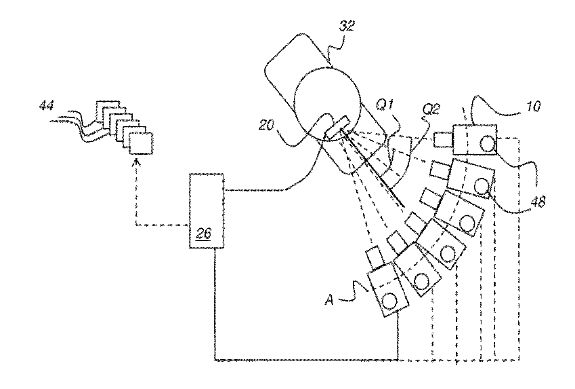
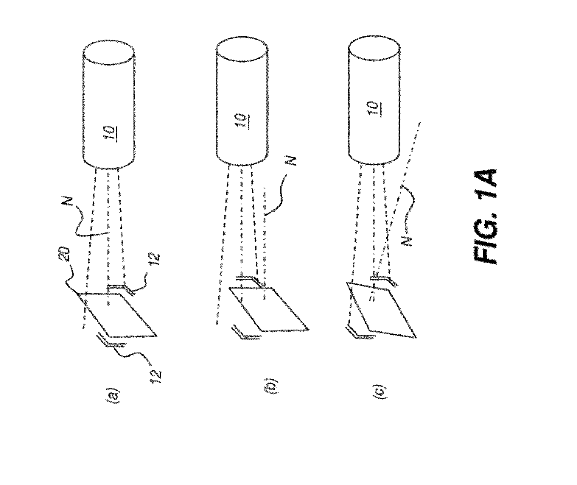
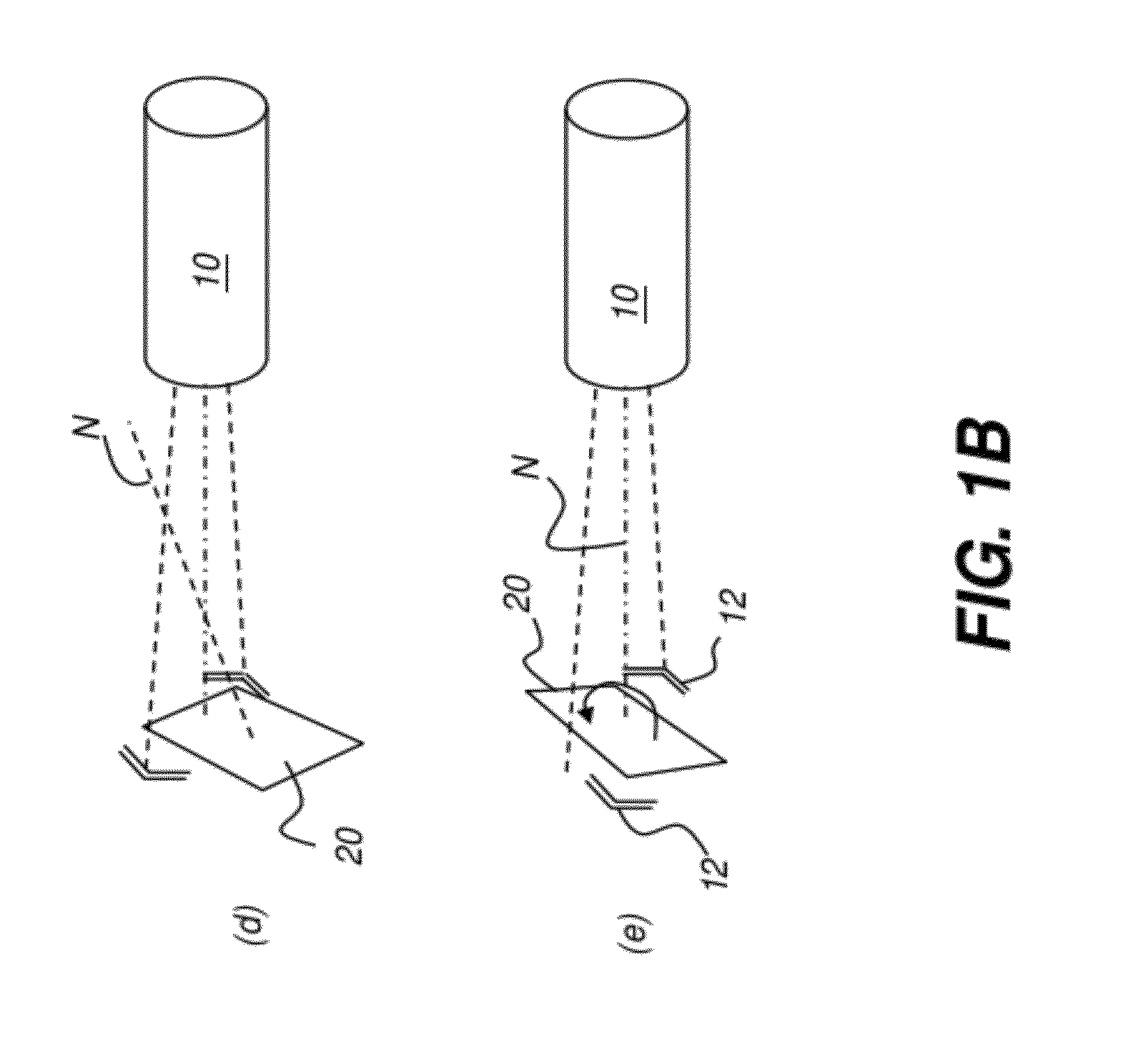
[0028]In the present disclosure, the term “detector” refers to the element that is placed in the patient's mouth, receives radiation, and provides the image content. Such a detector is a digital detector that provides the x-ray image data directly to an imaging system.

[0029]Detector alignment can be difficult for dental or intraoral radiography. The detector position is within the patient's mouth and is not visible to the technician. Instead, the technician typically places the detector into some type of holder, and then inserts the holder into place in the mouth. The holder may have a bite plate or other type of supporting member that helps to position the detector appropriately. Holders of this type can be cumbersome and uncomfortable to the patie...


PUM


Abstract

A method for obtaining an intraoral x-ray image determines an initial spatial position and angular orientation of an x-ray source relative to a detector. A first x-ray image is obtained with the x-ray source at the initial spatial position and angular orientation and stored, associating the initial spatial position and angular orientation to the first x-ray image. A sequence repeats that calculates a next spatial position and angular orientation for the x-ray source, provides positional adjustment information between the x-ray source and detector for obtaining a next x-ray image, measures and records the actual spatial position and angular orientation of the x-ray source relative to the detector and obtains and stores the next x-ray image at the measured spatial position and angular orientation, and forms a composite image using image data from the first and from the one or more next x-ray images.

Description

FIELD OF THE INVENTION[0001]The invention relates generally to diagnostic imaging and more particularly to an apparatus and method for obtaining multiple intraoral x-rays of a tooth to generate a 3D image representative of the tooth volume.BACKGROUND OF THE INVENTION[0002]A 3-dimensional (3-D) or volume x-ray image can be of value for diagnosis and treatment of teeth and supporting structures. A volume x-ray image for this purpose is formed by combining image data from two or more individual 2-D projection images, obtained within a short time of each other and with a well-defined angular and positional geometry between each projection image and the subject tooth and between each projection image and the other projection images. Cone-Beam Computerized Tomography (CBCT) is one established method for obtaining a volume image of dental structures from multiple projection images. In CBCT imaging, an image detector and a radiation source orbit a subject and obtain a series of x-ray projec...

Claims


Application Information

Patent Timeline
IPC IPC(8): A61B6/14A61B6/03
CPCA61B6/14A61B6/4085A61B6/466A61B6/5205A61B6/4494A61B6/4452A61B6/587A61B6/08A61B6/425A61B6/547
Inventor BOTHOREL, SYLVIESHELLARD, EDWARD R.INGLESE, JEAN-MARC
Owner CARESTREAM DENTAL TECH TOPCO LTD
Why Eureka
Try Eureka