Eureka AIR delivers breakthrough ideas for toughest innovation challenges, trusted by R&D personnel around the world.

System and method for discovering image quality information related to diagnostic imaging performance

a diagnostic imaging and image quality technology, applied in the field of system and method for discovering image quality information related to diagnostic imaging performance, can solve the problems of rarely retrieved data for other purposes, no attempt is made to systematically seek out such image quality information from within the digital diagnostic image stored in the vast storage bank of patient image data that is archived by hospitals and other health facilities, and achieves the effect of advance the art of healthcare administration

Inactive Publication Date: 2011-10-06
CARESTREAM HEALTH INC
View PDF1 Cites 31 Cited by
  • Summary
  • Abstract
  • Description
  • Claims
  • Application Information

AI Technical Summary

Benefits of technology

[0016]An object of the present invention is to address the shortfalls of existing data mining approaches for medical images and information and to advance the art of healthcare administration and delivery thereby.
[0025]An advantage provided by embodiments of the system of the present invention is that administrative information that spans multiple patient records, including patient images, can be obtained and analyzed for improving imaging performance.

Problems solved by technology

Once diagnosis is complete, the data stored is rarely retrieved for other purposes.
Once an image is stored, however, there is generally little likelihood of its data being utilized for any other purpose.
In conventional practice, however, no attempt is made to systematically seek out such image quality information from within the digital diagnostic image stored in the vast storage banks of patient image data that is archived by hospitals and other health facilities.
Their ability to detect visible defects and exercise proper judgement can be affected by factors such as difficulty in viewing images at the proper resolution and under the best possible conditions, demanding workloads, and varying levels of training and experience.
One or more of these factors can lead to defect oversight, so that images having marginal diagnostic quality at best may be stored in the PACS for use by the radiologist, without any short- or long-term correction taken.
Retaking the radiographic image, although it may be best for diagnostic accuracy, is highly undesirable for the patient and for efficient administration for a number of reasons.
This activity requires rescheduling complications, cost, and delays, and introduces other administrative problems.
As a result, some compromises can be made related to image quality, which can include accepting visually inspected images of disappointing quality in order to avoid huge disruptions in workflow, for example.
Without extensive effort, however, administrators and management personnel find it very difficult to uncover specific root causes of imaging problems that result in poor image quality and the need for retakes.
While such data mining techniques may be useful for obtaining comprehensive patient treatment data that is, of necessity, stored in various related systems, however, this diagnostic information relates only to each single patient, rather than to the performance of the imaging facility overall.
Thus, although data mining methods have been employed for obtaining information from different systems to aid in diagnosis of the individual patient, attention has not been paid to the particular difficulties and potential advantages of applying data mining techniques to information found within the diagnostic images themselves to produce image quality information for improved health care administration, particularly for improving image quality at a hospital or other diagnostic imaging site.

Method used


Image

  • System and method for discovering image quality information related to diagnostic imaging performance
  • System and method for discovering image quality information related to diagnostic imaging performance
  • System and method for discovering image quality information related to diagnostic imaging performance

Examples


example 1

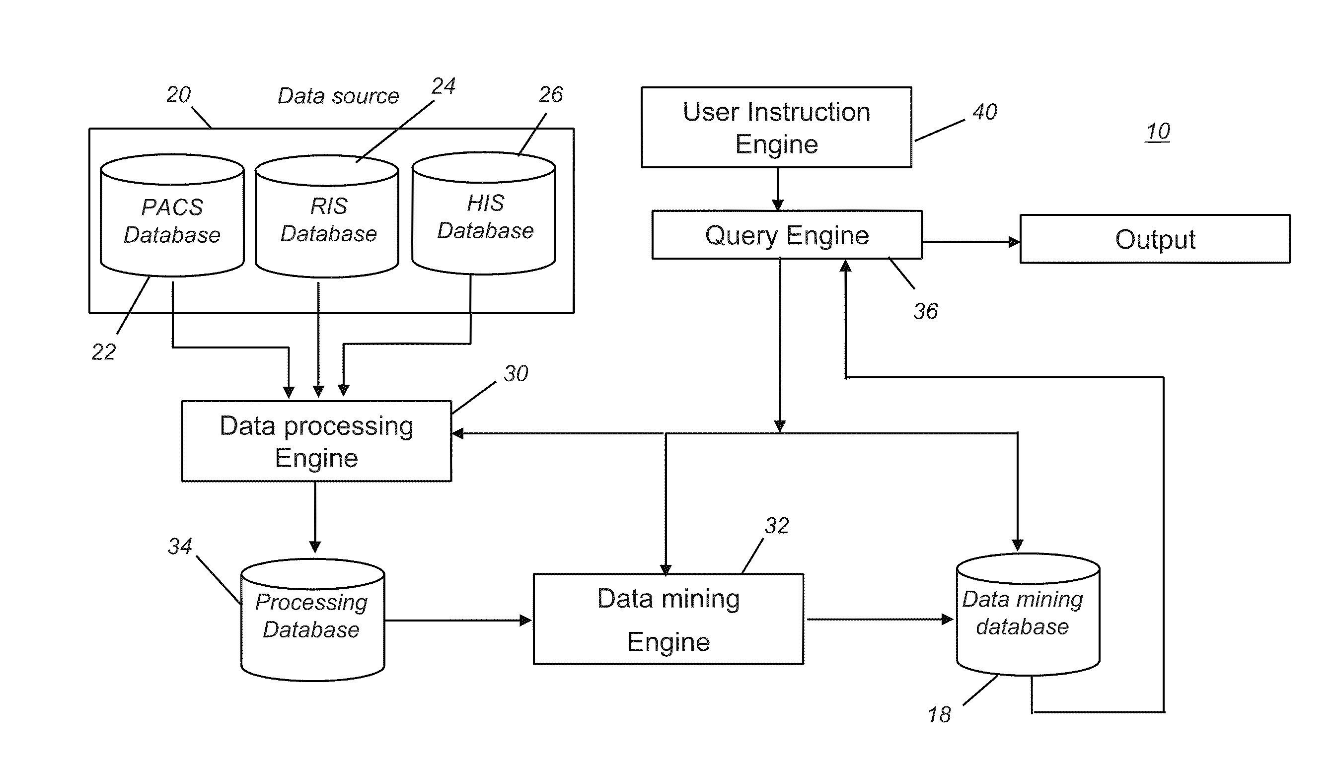
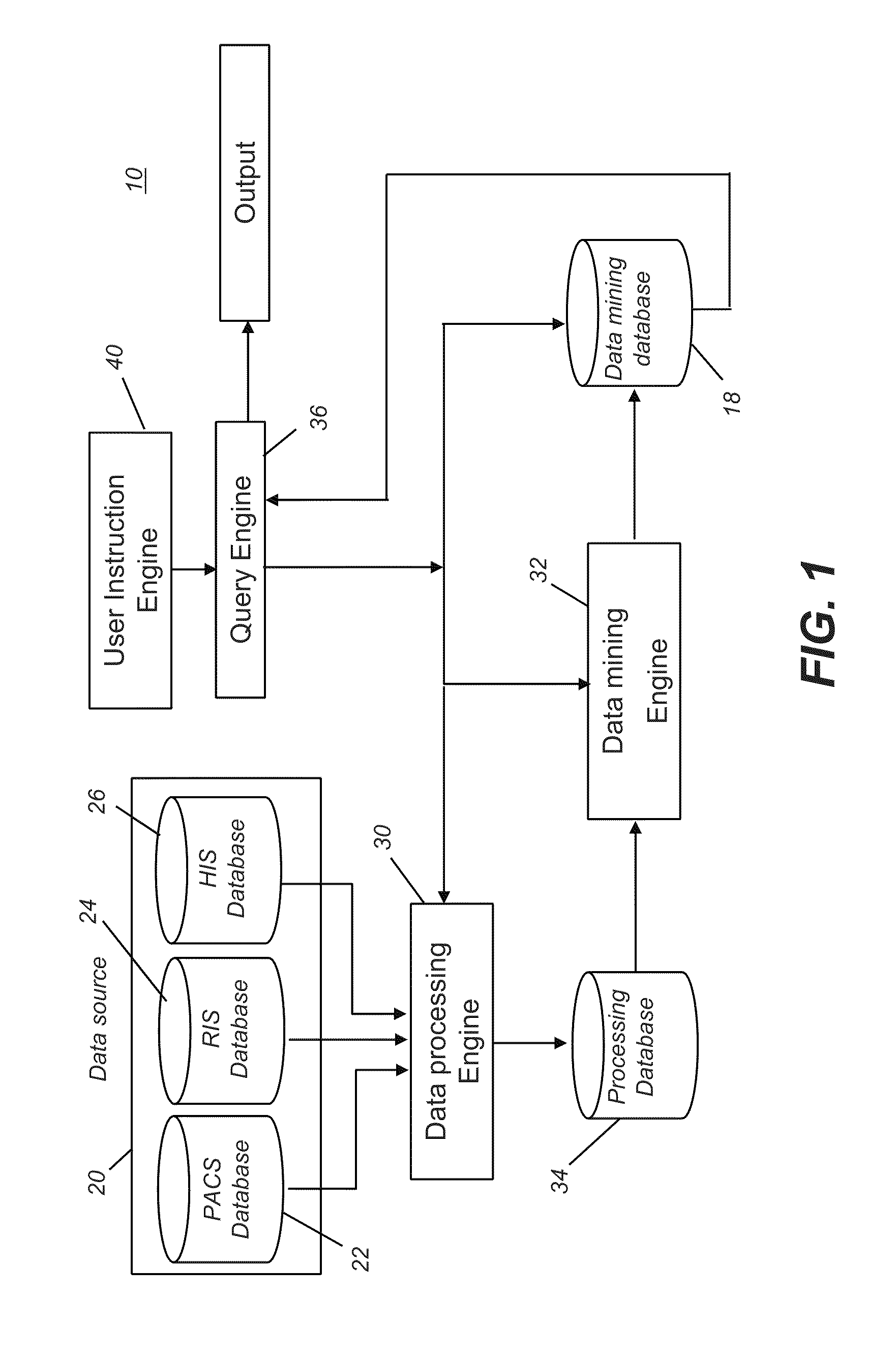
[0104]Administrators at a hospital are concerned with the amount of radiation that is used for chest imaging, with a goal to improving results obtained and eliminating radiation above a maximum threshold value. Periodic monitoring of these values is desired. To provide this information from data source 20 (FIG. 1), a technician using the system of the present invention makes the following GUI entries to user instruction engine 40:

Input Tab 94 Selections (FIG. 10A):

Mode:

Automated, BiWeekly

Location:

[0105]Softcopy interface

System Instructions Tab 96 Selections:

Data Processing Engine (FIG. 10B)

[0106]Exposure defects

Data Mining Engine (FIG. 10C)

[0107]Sum all exposure received by each study

Data Mining Database: (FIG. 10D)

[0108]Retrieve cumulative exposure data

Output Tab 98 Selections:

[0109]Format: Tabular, High level summary (FIG. 10E)

Locations: Hardcopy output, xyzz printer (FIG. 10F)

Ancillary actions: (none) (FIG. 10G)

[0110]The technician then initiates the database search process, base...

example 2

[0111]Due to excessive image quality defects reported by diagnosticians, training is recognized as a management priority for imaging personnel in a large medical facility. It is desirable to consider results from each imaging technician in order to help identify strengths and weaknesses and recommend additional training for individual technicians.

Input Tab 94 Selections: (FIG. 10A):

Mode:

Automated, BiWeekly

Location:

[0112]Softcopy interface

System Instructions Tab 96 Selections:

Data Processing Engine (FIG. 10B)

[0113]Clipped anatomy

Patient Motion

Artifacts

[0114]Exposure defects

Data Mining Engine (FIG. 10C)

[0115]Correlate image quality defects with technologist

Data Mining Database: (FIG. 10D)

[0116]Retrieve cumulative image quality data

Output Tab 98 Selections: (FIG. 10E)

Format: Graphical

[0117]Locations: Hardcopy output, xyzz printer (FIG. 10F)

Ancillary actions: (FIG. 10G)

Recommend training (condition #1, #2, etc.)

[0118]The technician then initiates the database search process, based on th...


PUM


Abstract

A system for discovering information related to diagnostic imaging performance at a medical imaging site. The system includes at least one database of stored digital diagnostic images; and a user instruction interface for obtaining an operator request for information related to image quality of the stored digital diagnostic images. A data processor is in communication with the at least one database, the data processor being programmed with instructions to use only information found within the stored digital diagnostic images themselves. A data mining engine is in communication with the data processor, the data mining engine being programmed with instructions to use only information found within the retrieved digital diagnostic images themselves.

Description

CROSS REFERENCE TO RELATED APPLICATIONS[0001]This application is a Continuation-in-Part of the following copending, commonly assigned, U.S. patent applications, the entire disclosures of which are incorporated by reference into this application:[0002](a) Ser. No. 12 / 190,613 filed Aug. 13, 2008 by Luo et al, entitled SYSTEM AND METHOD FOR DISCOVERING INFORMATION IN MEDICAL IMAGE DATABASE; and[0003](b) Ser. No. 11 / 834,304 filed on Aug. 6, 2007 by Luo et al, entitled METHOD FOR DETECTING ANATOMICAL BLUR IN DIAGNOSTIC IMAGES, now U.S. Pat. No. 7,899,229;[0004](c) Ser. No. 11 / 959,805 filed Dec. 19, 2007 by Wang et al, entitled SPECKLE REPORTING IN DIGITAL RADIOGRAPHIC IMAGING;[0005](d) Ser. No. 11 / 834,222 filed on Aug. 6, 2007 by Luo, entitled METHOD FOR DETECTING CLIPPED ANATOMY IN MEDICAL IMAGES, now U.S. Pat. No. 7,912,263; and[0006](e) Ser. No. 12 / 486,230 filed Jun. 17, 2009 by Wang et al, entitled AUTOMATED QUANTIFICATION OF DIGITAL RADIOGRAPHIC IMAGE QUALITY.FIELD OF THE INVENTION[...

Claims


Application Information

Patent Timeline
Patent Type & Authority Applications(United States)
IPC IPC(8): G06F17/30G16H30/20G16H30/40G16H50/20
CPCG06F19/3443G06F19/321G16H50/70G16H50/20G16H30/20G16H30/40
Inventor LUO, HUIWHALEY, JACQUELYN S.FOOS, DAVID H.
Owner CARESTREAM HEALTH INC
Why Eureka
Eureka Blog
Learn More