Laparoscopic wound closure device

a laparoscopic and wound technology, applied in the field of surgical suturing devices, can solve the problems of bowel strangulation or omental trapping, affecting the healing effect of the wound, so as to prevent post-operative herniation and reduce the occurrence of nerve entrapment.

US20160278763A1Active Publication Date: 2016-09-29MODERN SURGICAL SOLUTIONS
23 Cites 4 Cited by

Patent Information

Authority / Receiving Office
US · United States
Current Assignee / Owner
Publication Date
2016-09-29

Smart Images

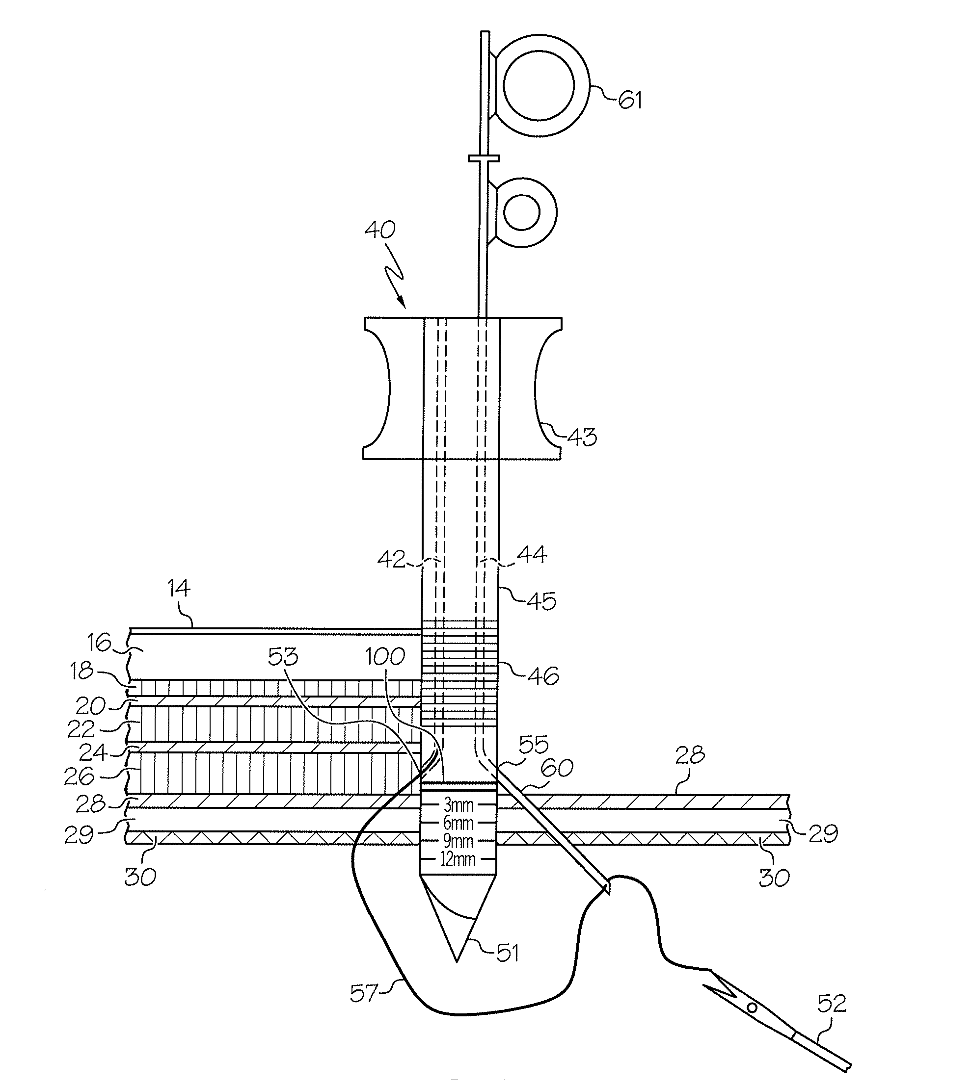
  • Figure 1
    Figure 1
  • Figure 2
    Figure 2
  • Figure 3
    Figure 3
Patent Text Reader

Abstract

A laparoscopic wound closure device includes an elongated body portion including a proximal end, a distal end, and a pair of vertical suture channels. Each vertical suture channel has an entrance at the proximal end and an exit near the distal end for passing a suture needle through, and travels parallel to the longitudinal axis of the body portion of the device. The vertical suture channels are designed to cause a suture needle to deflect laterally as it exits the wound closure device. The wound closure device can be an obturator or a sheath of a trocar assembly, and can be used to inject local anesthetic at the conclusion of surgery.
Need to check novelty before this filing date? Find Prior Art

Description

FIELD OF THE INVENTION

[0001] The present invention relates generally to surgical suturing devices, and more particularly to abdominal suturing devices designed for closing laparoscopic puncture wounds.BACKGROUND OF THE INVENTION

[0002] Laparoscopic surgery, or minimally invasive surgery, has replaced many standard invasive abdominal operations that require large incisions. Current laparoscopic surgical techniques include the creation of small incisions in the lateral abdominal wall, into which one or more surgical trocar assemblies or other similar puncturing devices are placed. A trocar assembly is typically comprised of an obturator having a puncturing distal end, and a sheath or cannula which is adapted to slide over the outer diameter of the obturator. The obturator and cannula are typically used together to puncture the lateral abdominal wall to gain access to the surgical site. Once the puncture is made, the obturator is removed from the hollow cannula creating a port for introdu...

Examples

Embodiment Construction

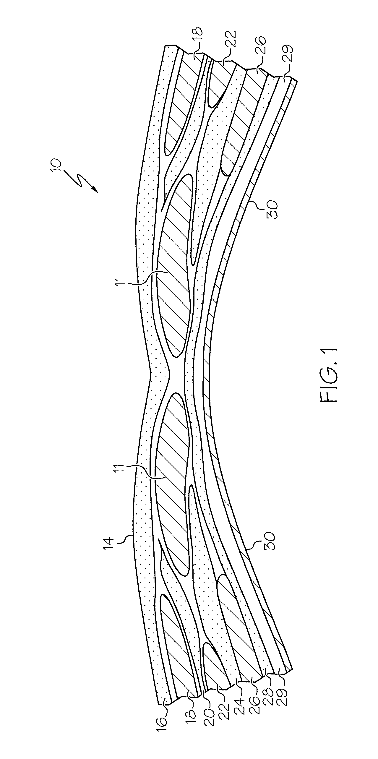
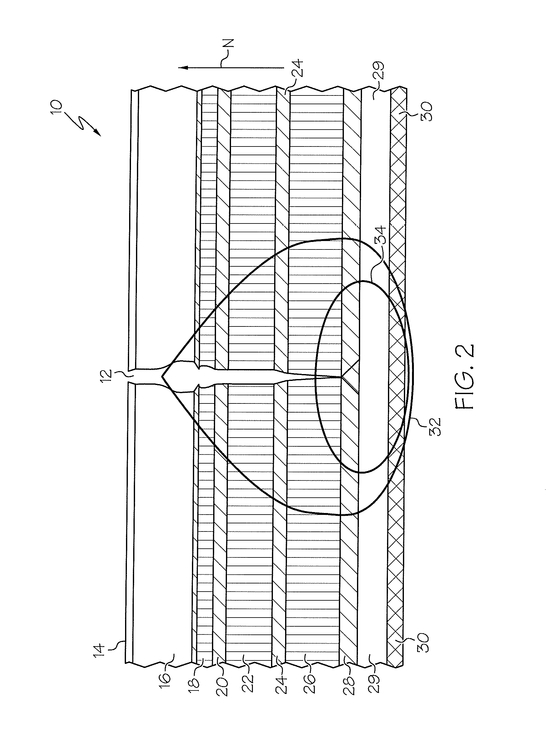
[0021]FIG. 1 illustrates the general anatomy of the abdominal wall 10, which is initially covered by a layer of skin 14 and fat 16 and composed of pairs of muscles, notably the rectus abdominis 11, the external oblique 18, the internal oblique 22, and the transversus abdominis 26 muscles. These muscles and their fascial attachments, notably the external oblique fascia 20, the internal oblique fascia 24 and the transversalis fascia 28, interdigitate and unite to form a sturdy, protective musculofascial layer that gives strength and support to the anterolateral abdominal wall. Below the level of the transversalis fascia 28, a layer of pre-peritoneal fat 29 precedes the peritoneum 30. The pre-peritoneal fat 29 is of varying thickness from patient to patient, ranging from zero mm to about 20 mm depending on factors such as the body fat index of the patient. The peritoneum 30 is a smooth, serous membrane that forms the lining of the intraperitoneal space within the abdominal cavity.

[0022...