
Device and Method for Assessment of Left Ventricular Ejection Fraction and Other Parameters of Cardiac Performance
a technology of ejection fraction and cardiac performance, applied in the direction of heart/pulse rate measurement, catheterization, sensor, etc., can solve the problems of insufficient quality of images to accurately assess ventricular volume, low correlation between ef obtained and mri, and inability to obtain images of sufficient quality
- Summary
- Abstract
- Description
- Claims
- Application Information
AI Technical Summary
Benefits of technology
Problems solved by technology
Method used
Image

Examples
Embodiment Construction
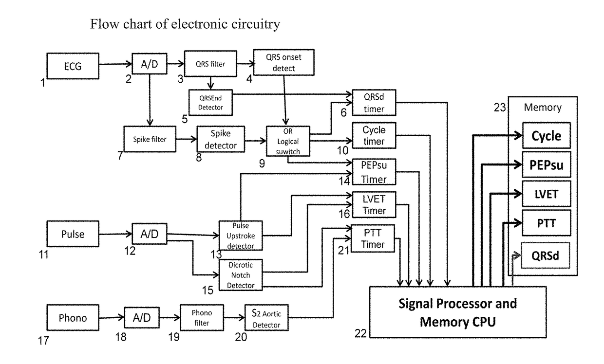
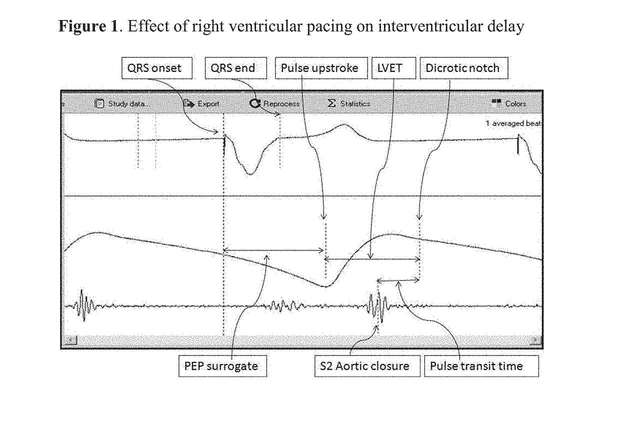
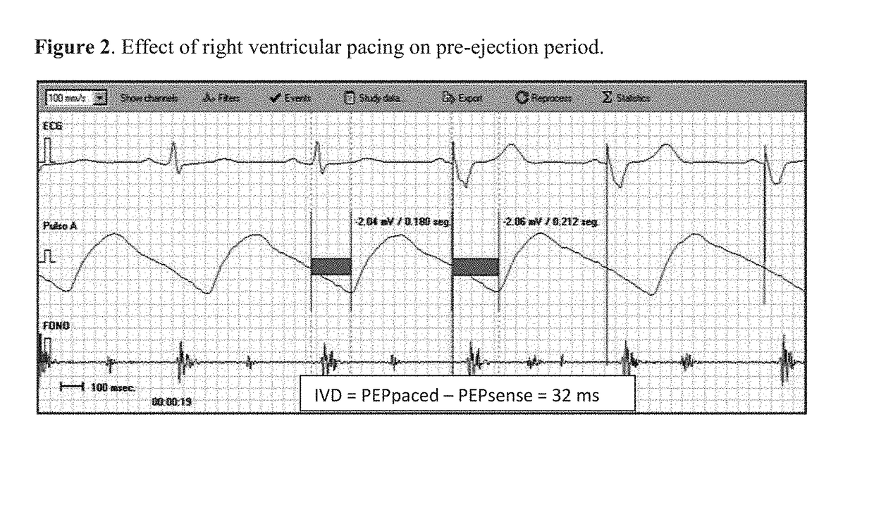
[0018]In a preferred embodiment of the invention, estimation of ejection fraction is done using a combination of physiologic parameters and dedicated software. Said physiologic parameters are the so-called systolic time intervals, obtained from a plurality of inputs, such as the ECG, an arterial pulse and a phonocardiogram. In this example, ECG is obtained from three standard disposable electrodes placed in a triangular fashion on the right clavicle, the right costal border and at the mid-clavicular line at the level of the 4th inter-costal space. The peripheral pulse is obtained with a commercially available transmittance O2 saturation sensor placed on a finger, although reflectance types are also suitable. The phonocardiogram, is recorded with a commercially available microphone, such as part # Part #: TSD108 from Biopac Systems Inc or similar. The tracings shown in FIGS. 1, 2 and 5 were obtained with the prototype at a sampling rate of 4 KHz for an accurate detection of pacemaker...
PUM
 Abstract
Description
Claims
Application Information