Distal anchor apparatus and methods for mitral valve repair

a technology of mitral valve and anchorage, which is applied in the direction of heart valves, annuloplasty rings, etc., can solve the problems of affecting the proper functioning of one or more of the valves of the heart, affecting the function of the valve in the heart, and prolapse and regurgitation of the valves

Active Publication Date: 2018-10-11
UNIV OF MARYLAND BALTIMORE +1
View PDF3 Cites 69 Cited by
  • Summary
  • Abstract
  • Description
  • Claims
  • Application Information

AI Technical Summary

Problems solved by technology

Various disease processes can impair the proper functioning of one or more of the valves of the heart.
Additionally, damage to the ventricle from prior heart attacks (e.g., myocardial infarction secondary to coronary artery disease) or other heart diseases (e.g., cardiomyopathy) can distort the geometry of the heart causing valves in the heart to dysfunction.
The vast majority of patients undergoing valve surgery, such as mitral valve surgery, suffer from a degenerative disease that causes a malfunction in a leaflet of the valve, which results in prolapse and regurgitation.
One possible malfunction, valve stenosis, occurs when a valve does not open completely and thereby causes an obstruction of blood flow.
Typically, stenosis results from buildup of calcified material on the leaflets of the valves causing them to thicken and thereby impairing their ability to fully open and permit adequate forward blood flow.
Another possible malfunction, valve regurgitation, occurs when the leaflets of the valve do not close completely thereby causing blood to leak back into the prior chamber.
The otherwise normally functioning leaflets do not have enough surface area to cover the enlarged orifice and fail to form a tight seal (e.g., do not coapt properly) causing regurgitation.
A Carpentier's type III malfunction involves restriction of the motion of one or more leaflets such that the leaflets are abnormally constrained below the level of the plane of the annulus.
MR results in a volume overload on the left ventricle which in turn progresses to ventricular dilation, decreased ejection performance, pulmonary hypertension, symptomatic congestive heart failure, atrial fibrillation, right ventricular dysfunction and eventually death.
The mitral valve and tricuspid valve, on the other hand, are more prone to deformation.
Deformation of the leaflets, as described above, prevents the valves from closing properly and allows for regurgitation or back flow of blood from the ventricle into the atrium, which results in valvular insufficiency.
However, because of the complex and technical demands of the repair procedures, the mitral valve is still replaced in approximately one third of all mitral valve operations performed in the United States.
Removing a portion of one or both of the mitral valve leaflets during such a resectional repair decreases the available leaflet tissue to seal the mitral orifice.
Implanting an annuloplasty ring introduces various risks.
Further, an annuloplasty ring can lead to infection and / or annuloplasty ring dehiscence: a well-documented failure mode of valve repair surgery.
Implanting an annuloplasty ring can interfere with such normal functioning of the heart.
Because the native leaflet tissue is maintained in non-resectional repairs, they often result in a larger surface of coaptation between the posterior and anterior mitral valve leaflets, but properly sizing the cords on a flaccid heart can be very challenging, especially for the low volume mitral valve surgeon.
Implanting an annuloplasty ring in a beating heart repair is technically challenging and rarely done in practice due in large part to the costs associated with two separate procedures (e.g., cordal repair and annuloplasty).
Unfortunately, there is no gold standard surgical treatment paradigm for functional MR and most functional MR patients are not referred for surgical intervention due to the significant morbidity, risk of complications and prolonged disability associated with cardiac surgery.
Surgeons use a variety of approaches ranging from valve replacement to insertion of an undersized mitral valve annuloplasty ring for patients suffering from functional MR and the long-term efficacy is still unclear.
According to some, a subsequent sub-analysis of subjects in the repair group suggests that the people who received a “good repair” did better than the replacement group but that when the repair arm was compared to mitral valve replacement, the “bad repairs” caused the replacement arm to perform better.
These procedures, however, adversely affect almost all of the organ systems of the body and may lead to complications, such as strokes, myocardial “stunning” or damage, respiratory failure, kidney failure, bleeding, generalized inflammation, and death.
The risk of these complications is directly related to the amount of time the heart is stopped (“cross-clamp time”) and the amount of time the subject is on the heart-lung machine (“pump time”).

Method used


Image

  • Distal anchor apparatus and methods for mitral valve repair
  • Distal anchor apparatus and methods for mitral valve repair
  • Distal anchor apparatus and methods for mitral valve repair

Examples


Embodiment Construction

[0122]The headings provided herein, if any, are for convenience only and do not necessarily affect the scope or meaning of the claimed invention.

Overview

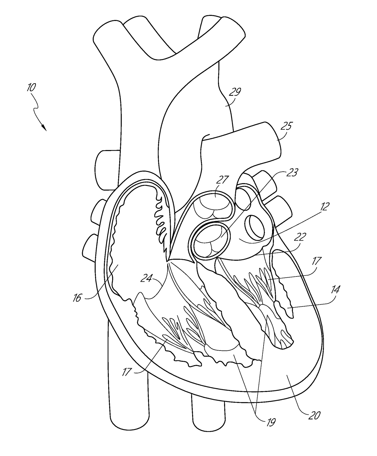
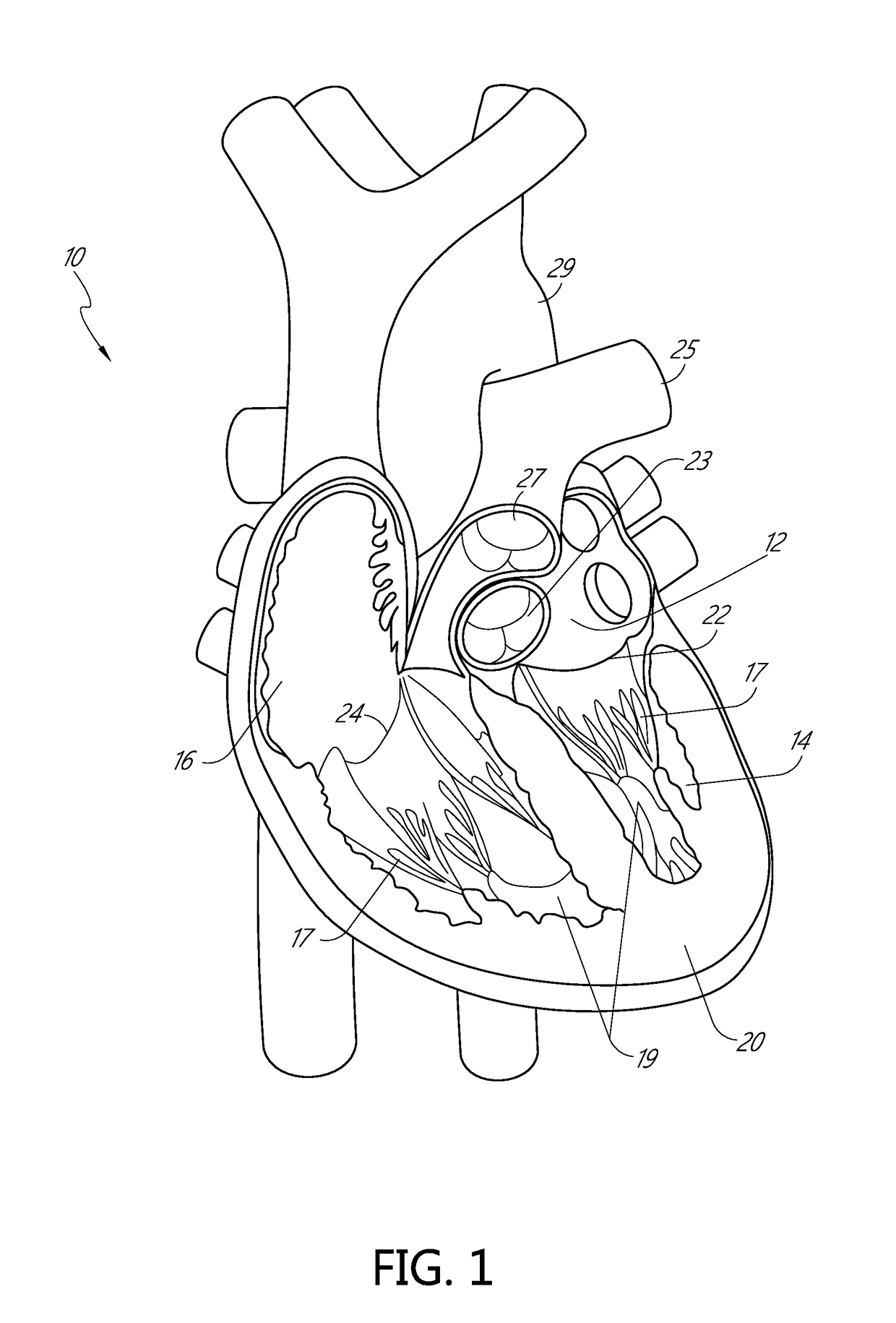
[0123]Apparatus and methods for performing a non-invasive procedure to repair a cardiac valve, such as a mitral valve or tricuspid valve, are described herein. In some embodiments, a method for repairing a mitral valve includes inserting a delivery device through an apex region (or adjacent to the apex region) of a heart and extending a distal end of the delivery device to the proximal side of a leaflet of the mitral valve. A piercing portion of the delivery device can be used to form an opening in the leaflet, through which the distal end of the delivery device can be inserted. The delivery device can be used to form or deliver a distal anchor to the distal side of the leaflet. The delivery device can then be withdrawn and a tether coupled to the distal anchor can be secured to an outer surface of the heart at the apex region with,...


PUM


Abstract

Described herein are devices and methods for mitral valve repair. The devices and methods implant a plurality of distal anchors at an annulus of the mitral valve (e.g., the posterior annulus) and tension artificial chordae to pull the portion of the annulus toward an opposite edge and inward into the ventricle. This can effectively reduce the size of the orifice and increase coaptation. The anchor points of the artificial chordae can be offset from the apex of the heart to achieve a targeted force vector on the annulus. In addition, some embodiments include distal anchors implanted in the posterior leaflet in addition to those at the posterior annulus. This allows differential tensioning to be applied to provide for greater flexibility in treating various configurations of mitral valves to improve coaptation.

Description

CROSS-REFERENCE TO RELATED APPLICATIONS[0001]This application is a continuation of International Patent Application No. PCT / US18 / 26570, filed Apr. 6, 2018, which claims the benefit of U.S. Application No. 62 / 482,468, filed Apr. 6, 2017, the contents all of which are expressly incorporated by reference herein in their entireties for all purposes.BACKGROUNDField[0002]Some embodiments described herein relate to methods and apparatus for performing cardiac valve repairs, and more particularly, methods and apparatus for performing minimally invasive mitral or tricuspid valve repairs.Description of Related Art[0003]Various disease processes can impair the proper functioning of one or more of the valves of the heart. These disease processes include degenerative processes (e.g., Barlow's Disease, fibroelastic deficiency), inflammatory processes (e.g., Rheumatic Heart Disease), and infectious processes (e.g., endocarditis). Additionally, damage to the ventricle from prior heart attacks (e.g....

Claims


Application Information

Patent Timeline
Patent Type & Authority Applications(United States)
IPC IPC(8): A61F2/24
CPCA61F2/2445A61F2/2451A61F2/2466A61F2/2457A61F2/2427A61F2/2412
Inventor D'AMBRA, MICHAEL NICHOLASCORTEZ, JR., FELINO V.GAMMIE, JAMES S.WILSON, PETEREPSTEIN, STEPHENCOURNANE, STEPHENETHERIDGE, JULIE MARIEBOYD, PETER
Owner UNIV OF MARYLAND BALTIMORE
Why Eureka
Try Eureka