Method for automatically setting an X-ray dosage for producing an X-ray tomographic image

a technology of x-ray tomography and automatic setting, which is applied in the field of automatic setting of x-ray dosage for producing a tomographic image, can solve the problems of inability to do x-ray dose, unavoidable noise in the image, and inability to do indiscriminately, and achieve the effect of constant image quality

Active Publication Date: 2006-05-02
SIEMENS HEALTHCARE GMBH
View PDF3 Cites 15 Cited by
  • Summary
  • Abstract
  • Description
  • Claims
  • Application Information

AI Technical Summary

Benefits of technology

[0007]It is an object of the present invention to provide a method for adjusting the X-ray dose in the production of a computed tomography image wherein an overall image with constant image quality is produced without overexposing the examination subject to X-rays.

Problems solved by technology

In the production of images of an examination subject in a computed tomography system, it is unavoidable that a certain level of noise will be present in the image.
In general, the level of noise can be reduced by increasing the tube current, and thereby increasing the X-ray dose, however, obviously this cannot be done indiscriminately because overexposure of the examination subject to X-rays must be avoided.
Particularly in computed tomography examinations wherein a larger portion of the body is scanned, which may encompass such differently dimensioned body regions, it is undesirable, from the point of view of image quality, to select one and only one tube current / X-ray dose for the entire scan, because the dose may be suitable for scanning one portion of the subject, but may not be suitable for scanning another portion.

Method used


Image

  • Method for automatically setting an X-ray dosage for producing an X-ray tomographic image
  • Method for automatically setting an X-ray dosage for producing an X-ray tomographic image
  • Method for automatically setting an X-ray dosage for producing an X-ray tomographic image

Examples


Embodiment Construction

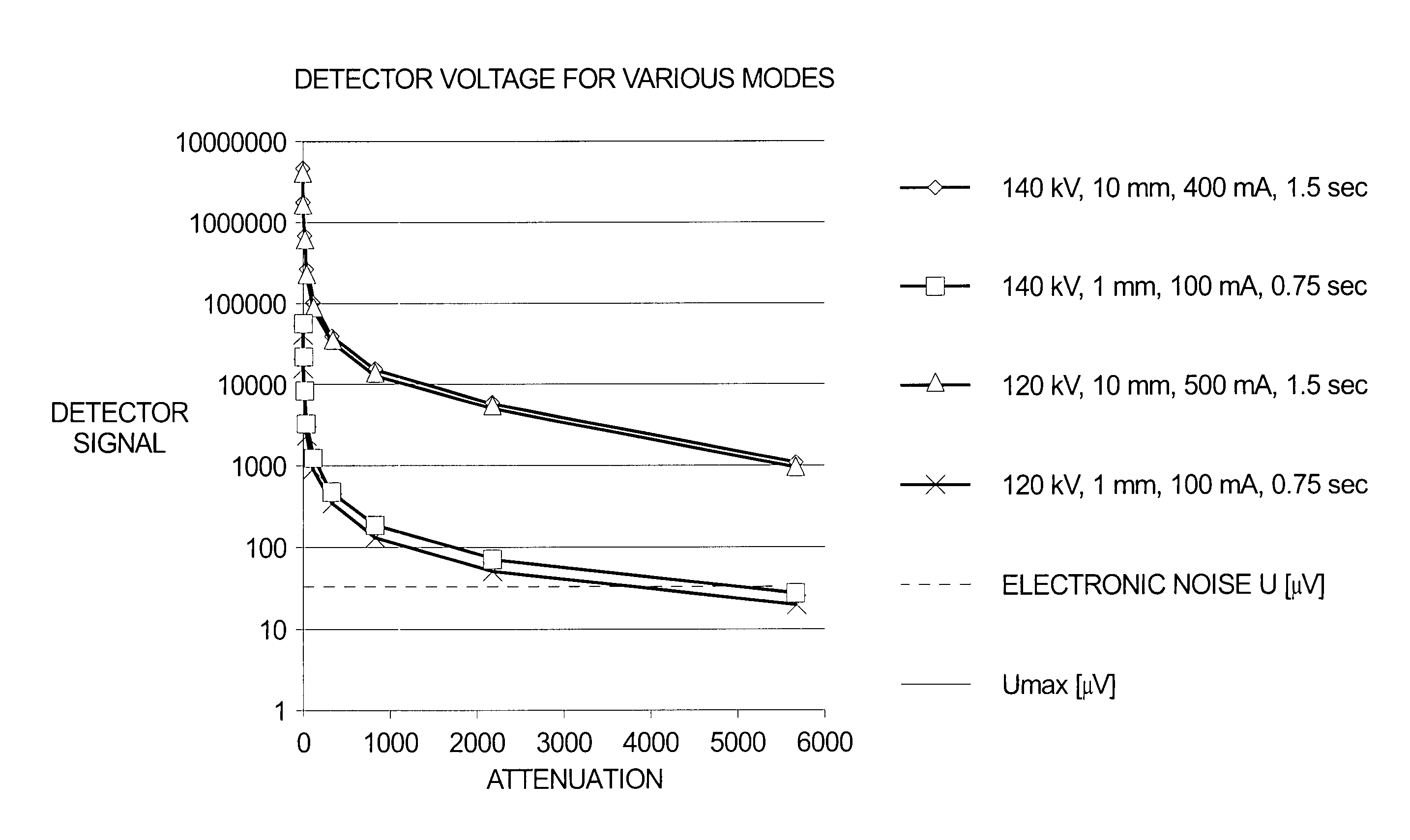
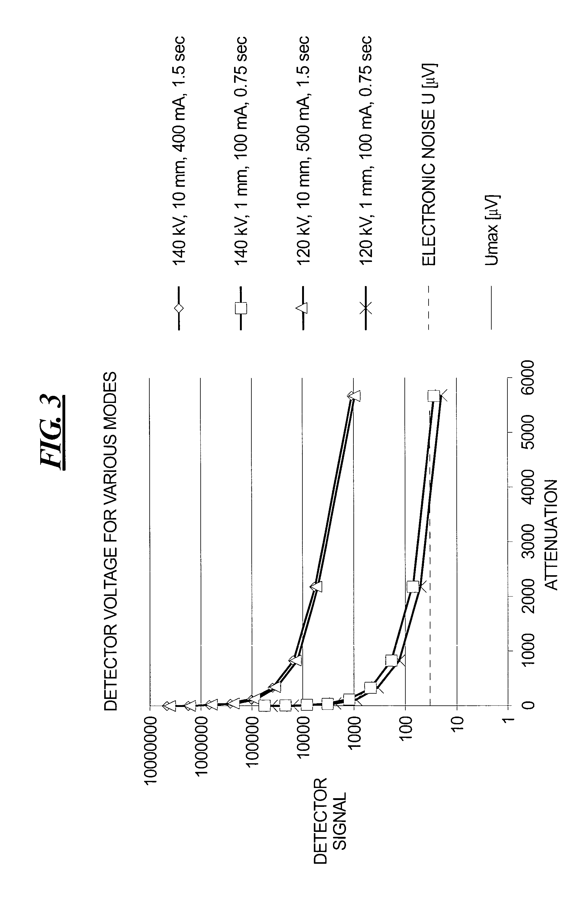
[0016]First the theoretical interrelation between measurement system, absorption and noise is summarized and is applied to measurements on the Plus 4. Subsequently, procedures for automatic dosage are discussed.

[0017]The number of quanta N0 emitted by the tube during the measurement of a projection and of the slice fade-in h depends on the tube high voltage kV, the tube current IRöhre, the pre-filtering V and the wedge filter W:

N0=N0(kV, IRöhre, h, V, W)  (1)

[0018]For a system as shown in FIG. 1, the pre-filtering can be assumed to be constant. The wedge filter causes a detector-channel-dependent variation of the quanta N0. The high voltage, as well as the pre-filtering and wedge filter, determines the spectral energy distribution of the x-ray spectrum.

[0019]For the functional interrelation of N with the high voltage U, the tube voltage I, and slice thickness h, the following holds:

[0020]N=N0*(UU0)2.26*(II0)*(hh0)(2)

It can be seen that N˜I, N˜h and N˜U2.26. If U=const, h=const, then...


PUM


Abstract

For automatically controlling or adjusting the X-ray dose when scanning an examination subject in a CT apparatus, a series of reference measurements are initially made by obtaining CT images of respective phantoms having different attenuation. The tube current for each phantom that produces an image of the phantom having an acceptable noise level, and thus an acceptable image quality, is stored in a table. In a subsequent CT scan of an examination subject, the tube voltage is automatically adjusted when irradiating a region of the subject having attenuation comparable to the attenuation of one of the phantoms, so as to employ the tube current when irradiating the examination subject that produced the image of acceptable quality for the comparable phantom.

Description

BACKGROUND OF THE INVENTION[0001]1. Field of the Invention[0002]The present invention is directed to a method for automatically setting an X-ray dosage for producing a tomographic image, using an X-ray CT device.[0003]2. Description of the Prior Art[0004]In the production of images of an examination subject in a computed tomography system, it is unavoidable that a certain level of noise will be present in the image. In general, the level of noise can be reduced by increasing the tube current, and thereby increasing the X-ray dose, however, obviously this cannot be done indiscriminately because overexposure of the examination subject to X-rays must be avoided.[0005]Moreover, different portions of a human examination subject exhibit different attenuations, due to respectively different compositions and physical dimensions. Particularly in computed tomography examinations wherein a larger portion of the body is scanned, which may encompass such differently dimensioned body regions, it ...

Claims


Application Information

Patent Timeline
Patent Type & Authority Patents(United States)
IPC IPC(8): H05G1/32A61B6/03A61B6/00H05G1/34H05G1/50
CPCA61B6/032A61B6/583A61B6/542A61B6/4035
Inventor POPESCU, STEFANSUB, CHRISTOPHWOLF, HEIKOHENTSCHEL, DIETMAR
Owner SIEMENS HEALTHCARE GMBH
Why Eureka
Try Eureka