Method and apparatus for acquisition and evaluation of image data of an examination subject

an examination subject and image data technology, applied in the field of acquisition and evaluation of image data of examination subjects, can solve the problems of insufficient production, inability to arrange or inability to compare exposures produced with different apparatuses, etc., to achieve fast and qualitatively better evaluation

Inactive Publication Date: 2008-12-16
SIEMENS HEALTHCARE GMBH
View PDF16 Cites 8 Cited by
  • Summary
  • Abstract
  • Description
  • Claims
  • Application Information

AI Technical Summary

Benefits of technology

[0007]An object of the present invention is to provide a method for acquisition and evaluation of image data of an examination subject that are obtained with at least one medical imaging apparatus, which allows a faster and qualitatively better evaluation compared to conventional methods.
[0017]The inventive method thus offers the possibility to quickly compare examination images of examinations that have occurred at different points in time or were implemented with different apparatuses using the spatial arrangement in the whole-body coordinate system. Through the knowledge of the associated coordinates of the whole-body coordinate system, it is immediately clear where the respective image is to be arranged in the patient body and how it compares with earlier exposures for evaluation. If a doctor or technician who is occupied with the evaluation of the image data searches for exposures of specific regions, he or she can simply search through a data processing device by specification of the coordinates of the whole-body coordinate system. Like the examination planning, the navigation through the (often large) quantity of image data is significantly simplified; reference to a single coordinate system that can be maintained unchanged for all examinations of the respective patient now suffices. The quality of the coverage thus can be distinctly improved while at the same time the examination planning (for example given the production of real-time exposures) is simplified.
[0022]The whole-body overview image and / or partial images (that, for example, were created in the framework of different examinations) can be mapped to one another, particularly by means of an iterative error correction method. Initially only rough correlations and deviations are determined, for example in order to implement a size adaptation, whereupon corrections can then be implemented step-by-step using specific image information. Such a superimposition of the whole-body overview images or partial images that were produced at various points in time enables errors to be avoided in the arrangement of the examination images in the original whole-body coordinate system, since inconsistencies as well as fundamental differences (for example with regard to the orientation) can be detected and accounted for with the mapping of the overview images or partial images to one another.
[0028]The examination images additionally can be arranged in an examination-based coordinate system. For example, in the case of a magnetic resonance examination such an examination-based coordinate system is based on the position of the magnet and is advantageous for monitoring the positioning of the produced images with regard to this specific examination apparatus. An additional spatial reference possibility results by the use of such an examination-based coordinate system, whereby the possibilities for examination evaluation are further improved.

Problems solved by technology

It is thereby frequently not sufficient to produce an exposure or an exposure series once with an examination apparatus.
In the use of apparatus-specific coordinate systems, there is the problem of the comparability of exposures that have been produced with different apparatuses.
With region-related patient coordinate systems, there is the problem that exposures of other body regions or exposures with different orientations cannot be arranged or compared, or can only be arranged or compared with difficulty.
It is extraordinarily difficult to interpret exposures that were produced with different apparatuses or at different times, or of different examination regions, in the evaluation and the subsequent finding.

Method used


Image

  • Method and apparatus for acquisition and evaluation of image data of an examination subject
  • Method and apparatus for acquisition and evaluation of image data of an examination subject
  • Method and apparatus for acquisition and evaluation of image data of an examination subject

Examples


Embodiment Construction

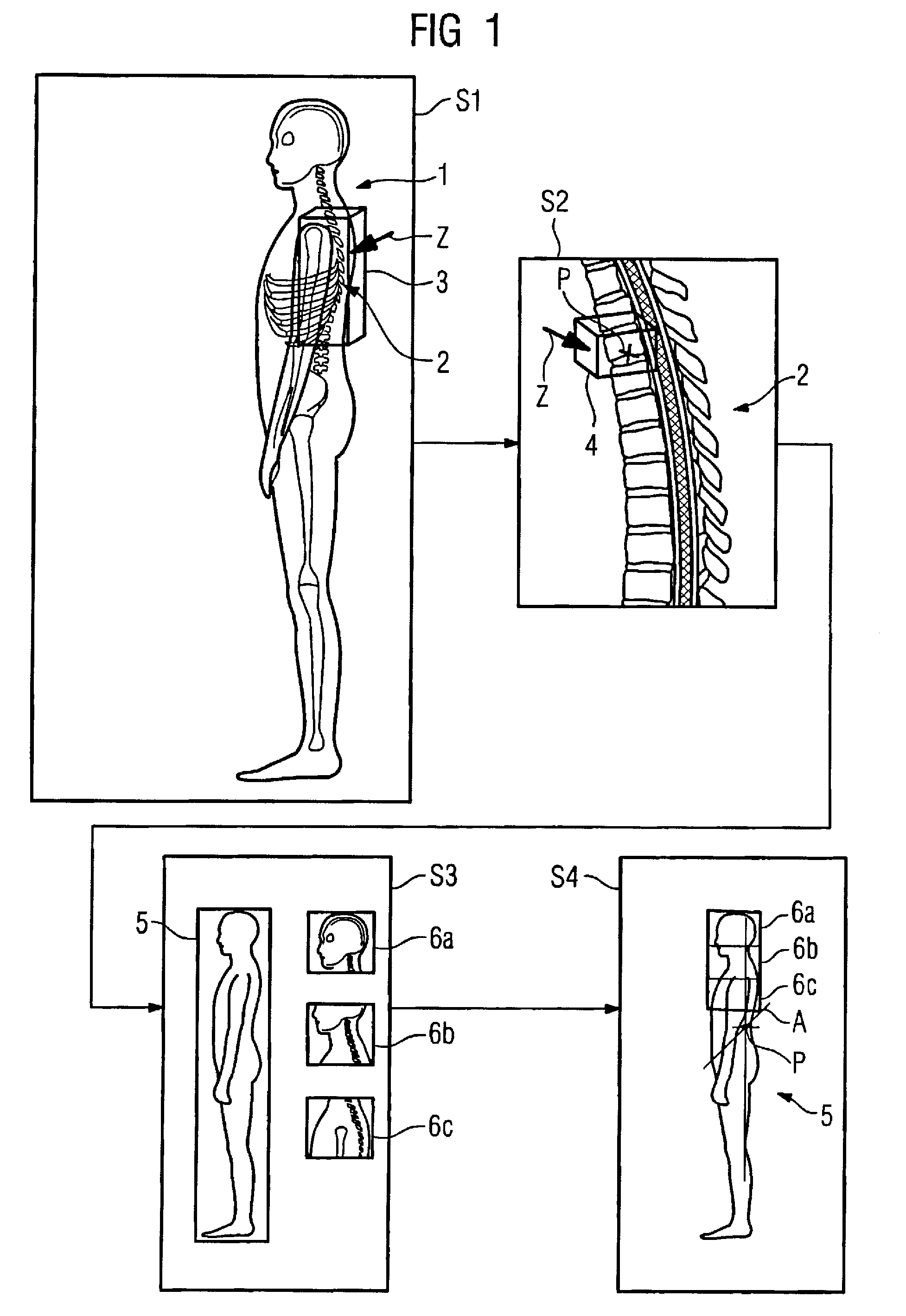
[0037]FIG. 1 shows workflow diagrams of an embodiment of the inventive method. In step S1, a whole-body overview image 1 of the examination subject has been initially created with an imaging medical examination apparatus, in which whole-body overview image 1 bone structures are indicated in order to indicate that it is an exposure obtained with a medical imaging apparatus. The overview image in this example shows a lateral view that is based on a three-dimensional data set. Alternatively, it is possible to use two-dimensional overview images that are based, for example, on exposures in a specific slice plane.

[0038]In the whole-body overview image 1, the doctor or medical assistant who operates the examination apparatus selects an anatomical region with the mouse point Z, by drawing a selection box 3 with the mouse pointer Z by means of an image processing program. In step S2, the selected anatomical region 2 (which here corresponds to a segment of the spinal column) is shown enlarge...


PUM


Abstract

In a method and apparatus for acquisition and evaluation of image data of an examination subject that are to be created with at least one imaging medical imaging apparatus, generation of a whole-body overview image of the examination subject is generated and selection of an anatomical region in the whole-body overview image is selected. A point in the selected anatomical region is established that forms the origin of a whole-body coordinate system relating to the examination subject, and establishment of an associated, subject-related coordinate system is established / Upon a later examination with a medical imaging apparatus, a whole-body overview image and / or a partial image containing the origin and / or another marker point is / are generated, and examination (diagnostic) images of the examination subject (S3), are generated. Using new whole-body overview images and / or the partial image, the examination images and spatially arranged in the original whole-body coordinate system dependent on a comparison of at least one item of image information of the examination image with at least one item of image information of the whole-body overview image and / or at least one item of information of the partial image that is related to the whole-body coordinate system.

Description

BACKGROUND OF THE INVENTION[0001]1. Field of the Invention[0002]The present invention concerns a method for acquisition and evaluation of image data of an examination subject that are generated with at least one medical imaging apparatus as well as an associated apparatus.[0003]2. Description of the Prior Art[0004]In the framework of the examination of a patient, exposures are often produced with medical imaging apparatuses in order, for example, to generate a diagnosis or a finding or to be able to plan a therapy. It is thereby frequently not sufficient to produce an exposure or an exposure series once with an examination apparatus. Rather, exposures are often acquired at specific time intervals, for example to monitor a recovery or an etiopathology (course of a disease), possibly also exposures of other body regions insofar as new diseases could occur or interactions are to be monitored.[0005]An examination with different examination apparatuses (for example with a computed tomogr...

Claims


Application Information

Patent Timeline
Patent Type & Authority Patents(United States)
IPC IPC(8): G06K9/00
CPCA61B6/00A61B6/463A61B6/469A61B6/5229A61B6/5241
Inventor HAIDER, SULTANKREISLER, PETER
Owner SIEMENS HEALTHCARE GMBH
Why Eureka
Try Eureka