Multi-modality medical data three-dimensional visualization method

A multimodal, volumetric data technology, applied in the field of biomedical engineering, can solve problems such as multimodal data fusion and display problems, and achieve the effect of avoiding falling into local extremum, increasing speed, and increasing registration speed

Inactive Publication Date: 2009-06-10
HANGZHOU DIANZI UNIV
View PDF12 Cites 1 Cited by
  • Summary
  • Abstract
  • Description
  • Claims
  • Application Information

AI Technical Summary

Problems solved by technology

The visualization of volume-volume fusion (03805385.3) proposes a method of using multiple volumes to fuse and display images. This invention uses the extraction of two-dimensional layers in the fusion display of three-dimensional volume data, and its essence is based on two-dimensional-two-dimensional fusion display method, did not solve the problem of real multi-modal data fusion display based on three-dimensional stereo

Method used


Image

  • Multi-modality medical data three-dimensional visualization method
  • Multi-modality medical data three-dimensional visualization method
  • Multi-modality medical data three-dimensional visualization method

Examples


Embodiment Construction

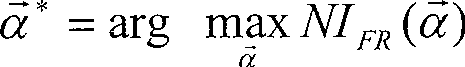
[0053] We illustrate the 3D visualization of CT and PET modal data with examples. Let the CT volume data be the reference volume, the PET volume data be the floating volume, and their respective coordinates be the object space coordinates. First, we set the multi-resolution coarse registration to 8 pixel length units, perform coordinate transformation on CT and PET data, and calculate the standardized mutual information after the coordinate transformation, and find the coordinate parameters when the standardized mutual information is the largest , take the optimal point as the starting point, reduce the range of coordinate transformation, and at the same time halve the unit length of coordinate transformation, that is, increase the resolution, continue the previous process of coordinate transformation and standardized mutual information criterion, until the optimal point, in the new minimum On the basis of optimization, repeat the previous steps, and approach step by step fro...


PUM


Abstract

The invention involves a multi-mode medical body data three-dimensional video-method. Most the existing technologies are based on two-two-dimensional lay or two-three dimensional lay, which do not realize the integrated display course of medical body data aligning and amalgamation. The said method includes standardization mutual information-way aligning based on multi resolution ratio and single drop multi-mode straight body protracting-way amalgamation display. The previous one includes three approaches, such as coordinate transform, orientation criterion and multi resolution ratio optimize; the latter includes five approaches, such as impress function definition, the calculation of illumination model, image composition, single drop multi-mode show of the multi-mode data and amalgamation display. The invention can realize the integrated display course of medical body data aligning and amalgamation, and is a real two-three dimensional multi-mode video-method.

Description

technical field [0001] The invention belongs to the field of biomedical engineering and relates to a three-dimensional visualization method for multimodal medical body data. Background technique [0002] Medical image data can be divided into two categories: one is anatomical images such as X-ray computed tomography (CT) and nuclear magnetic resonance (MR), which are suitable for displaying the anatomical details of the human body and have high spatial resolution; the other is normal Electron emission tomography (PET), single photon emission tomography (SPECT) and other functional images are suitable for reflecting information such as functional changes and metabolic abnormalities of human tissues, but their spatial resolution is poor. Due to the limitations of image information caused by different imaging principles, the effect of using one modality data alone is not ideal. Through multi-modal visualization, multiple scan sources or data from the same patient or multiple p...

Claims


Application Information

Patent Timeline
Patent Type & Authority Patents(China)
IPC IPC(8): G06T11/00G06T7/00
Inventor 金朝阳薛安克王建中杨成忠
Owner HANGZHOU DIANZI UNIV
Why Eureka
Try Eureka