3-D high-definition mammary gland imager

A high-definition breast imaging technology, applied in the field of biomedical imaging, can solve the problems of high clinical misdiagnosis rate and missed diagnosis rate, difficulty in judging the shape and location, and blurred shape of lesion tissue, so as to improve the signal-to-noise ratio and resolution, The effect of reduced volume and exquisite instrument structure design

Inactive Publication Date: 2007-08-15
BEIJING INSTITUTE OF TECHNOLOGYGY +1
View PDF0 Cites 15 Cited by
  • Summary
  • Abstract
  • Description
  • Claims
  • Application Information

AI Technical Summary

Problems solved by technology

However, the red light transmission imager generates a direct transmission image of the entire breast tissue, all shadows overlap, the resolution is low, the shape of the lesion tissue is blurred, and it is difficult to judge the specific shape and position, resulting in a high rate of clinical misdiagnosis and missed diagnosis

Method used


Image

  • 3-D high-definition mammary gland imager

Examples


Embodiment Construction

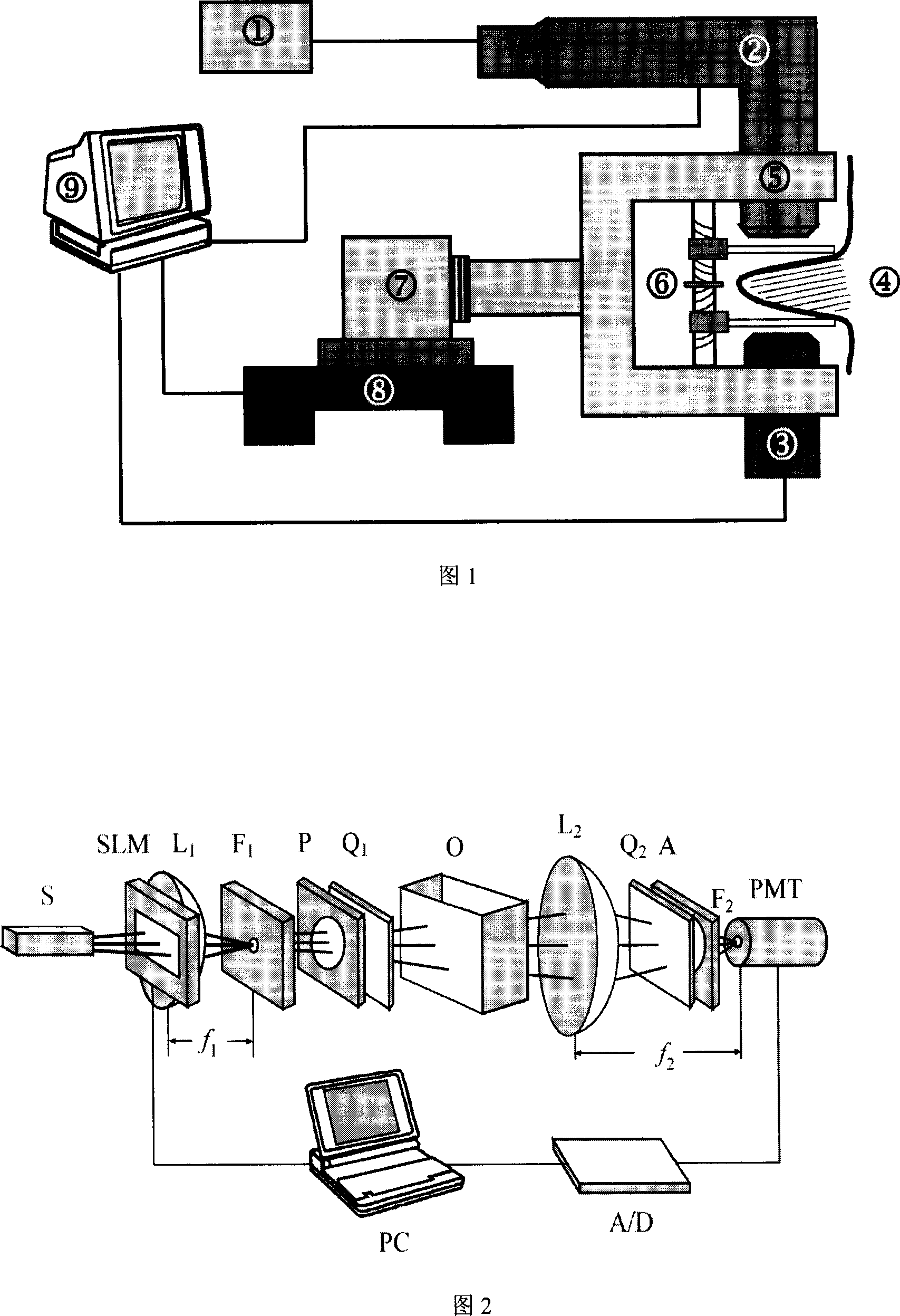
[0026] The present invention adopts non-coherent coded aperture imaging technology. Strong background noise is the common principle noise of this technology. We adopt the following methods to eliminate the inherent noise of this technology.

[0027] Method 1: The method of constructing a complementary hologram. Assume that the optimized strip plate transmittance function is F OZP , when it is displayed with a spatial light modulator, a DC term needs to be added, and this DC term is the factor that causes the background noise. First construct a positive encoding function (1+F OZP ) and a negative encoding function (1-F OZP ), and then subtract the positive and negative holograms obtained by scanning the two encoding functions respectively to obtain a complementary hologram. This removes background noise on the hologram due to the DC term. Use F when decoding OZP The complementary hologram is decoded to obtain a reconstructed image without background noise.

[0028] Method...


PUM


Abstract

The invention discloses a three-dimensional high-definition mammary gland imager in the biological medical imaging domain, which is characterized by the following: judging pathologic position and degree of mammary gland tissue according to optical parameter according to grey, tissue structure and size semi-quantitatively or qualitatively; adopting infrared (600-900nm) as light source; modulating space of irradiated light through coded aperture without harmful detecting; forming digital infrared optical scanning hologram; utilizing digital decoded technique to extract the information of galactophore tissue; displaying three-dimensional high-definition image.

Description

Technical field [0001] The invention belongs to the field of biomedical imaging, and is mainly used for the detection of breast diseases. By means of imaging, the internal structure of the breast can be displayed in a three-dimensional and high-definition manner. Quantitatively or qualitatively judge the location and degree of breast tissue lesions. Background technique [0002] At present, the methods used for breast disease examination are mainly various imaging methods, such as: mammography X-ray imaging, nuclear magnetic resonance imaging, ultrasound imaging, and infrared imaging. However, these traditional imaging techniques have certain limitations. Molybdenum target X-ray photography will cause ionizing radiation to the human body, damage the DNA in cells, and induce cell canceration. Although X-ray imaging has a high resolution, the disadvantages of destructive testing limit the general survey application of this imaging technology; the outstanding advantages of nu...

Claims


Application Information

Patent Timeline
Patent Type & Authority Applications(China)
IPC IPC(8): A61B5/00A61B19/00
Inventor 谢敬辉孙萍莫晓丽
Owner BEIJING INSTITUTE OF TECHNOLOGYGY
Why Eureka
Try Eureka